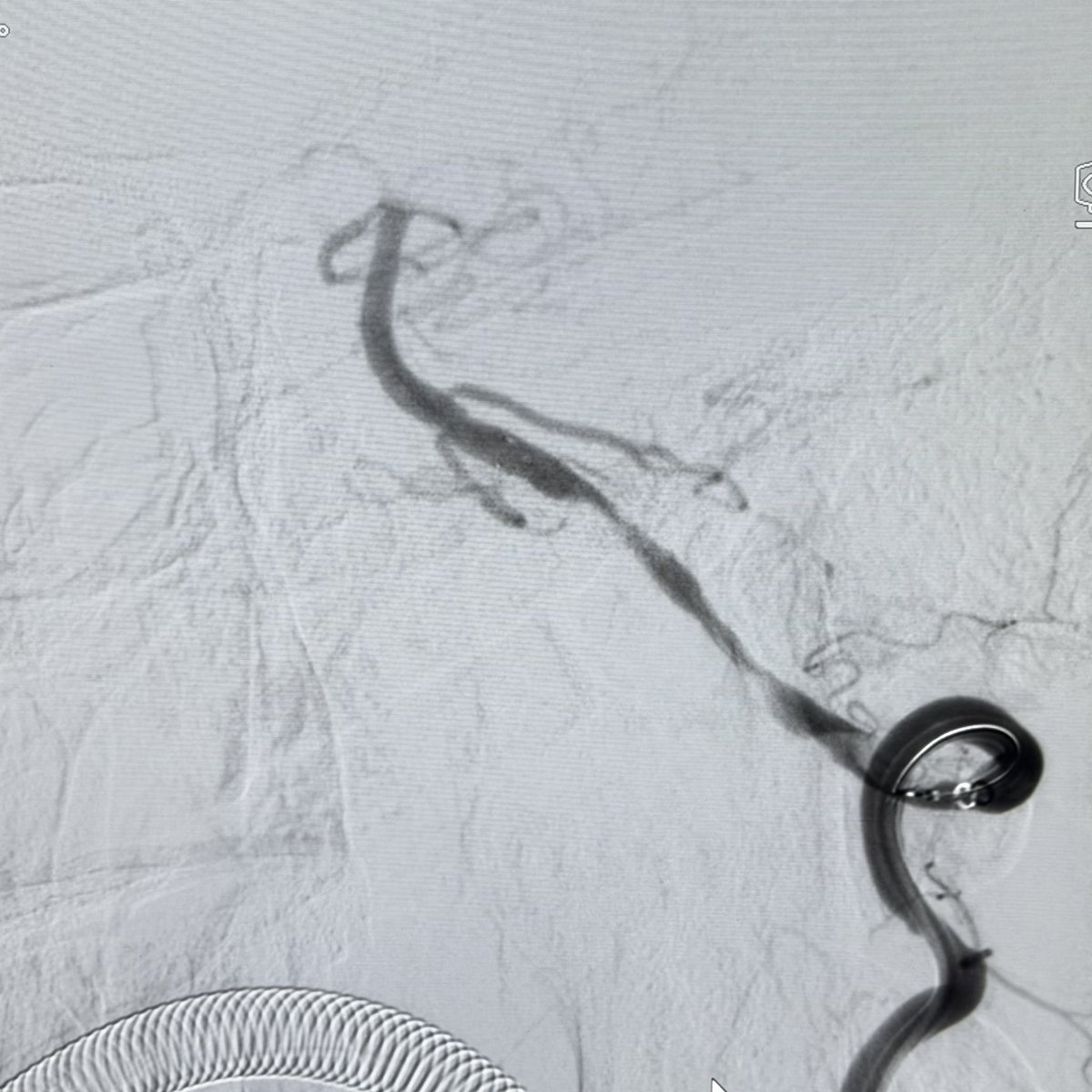
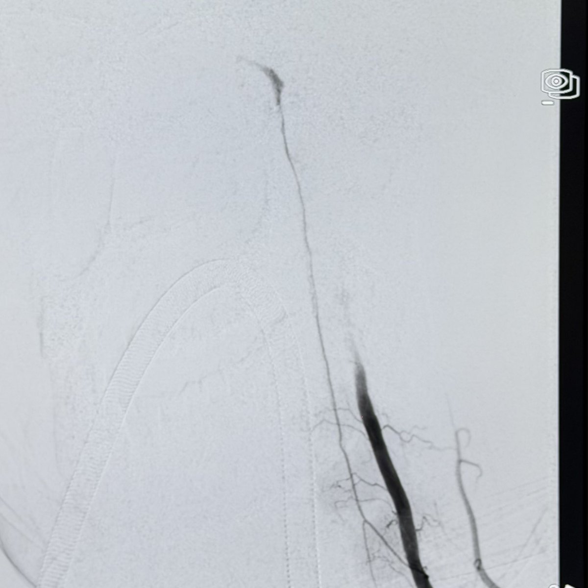
One pregnancy. Two babies. Two completely different outcomes.
What happened to these twins... and how can one baby be born healthy while the other is critically ill despite sharing the same womb?
🧠 What's the diagnosis? #MedTwitter#Neonatology
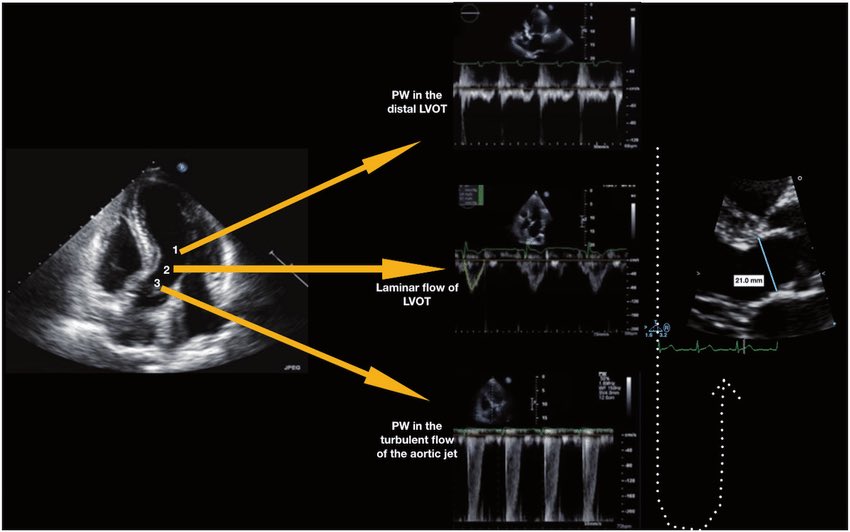
🫀 Did you know where the recommendation to place the pulsed wave Doppler sample volume 0.5–1 cm from the aortic valve to measure LVOT VTI comes from?
The answer is more interesting than it seems. It doesn’t come from a single study or an experiment designed for that purpose. It comes from a historical chain spanning nearly 40 years:
🔬 1982–1984 — The physical foundation
Pasipoularides and Murgo demonstrated using invasive catheters and mathematical models that in aortic stenosis there is a real zone of flow acceleration in the LVOT, just proximal to the valve, without any second anatomic obstruction. Pure hemodynamics — no Doppler yet.
👉 Bird et al. Circulation 1982 → https://t.co/oCXGNgWsPF
👉 Pasipoularides et al. Am J Physiol 1984 → https://t.co/cfvNMpEgzG
📐 1984 — The apical 5-chamber view
Lewis, Kuo and Quinones were the first to validate cardiac output measurement using pulsed wave Doppler from the cardiac apex. They described placing the sample volume “immediately proximal to the aortic valve leaflets” — but without specifying any distance in centimeters.
👉 Lewis et al. Circulation 1984 → https://t.co/lRVjLuYMEs
📏 1985 — The first numerical distance
Skjaerpe, Hegrenaes and Hatle (the Norwegian group) were the first to quantify this in Doppler: they empirically observed that flow acceleration began 0.5 to 1.5 cm proximal to the valve, and placed the sample volume just proximal to that zone. They directly cited Pasipoularides as supporting evidence. This was the first time a numerical distance appeared in the technique.
👉 Skjaerpe et al. Circulation 1985 → https://t.co/rf7l2FQUs4
📊 1986–1988 — Practical consolidation
Otto et al. used ~1.0 cm. Oh, Tajik and the Mayo Clinic group explicitly established the range of 0.5 to 1.0 cm in 100 patients, justifying it as necessary to avoid the subvalvular acceleration zone. This is the figure we all recognize today.
👉 Otto et al. JACC 1986 → https://t.co/avke1qOy4x
👉 Zoghbi et al. Circulation 1986 → https://t.co/KFiChfBNuc
👉 Oh et al. JACC 1988 → https://t.co/1C8sHRSMPq
📋 2002 — It becomes “official”
Quinones, Otto, Zoghbi and colleagues codified it in the ASE guidelines as “~5 mm proximal to the aortic valve”… but without citing any specific study to support it. It had already become expert consensus.
👉 Quiñones et al. JASE 2002 → https://t.co/U3hXFns4jx
⚔️ 2017 — The debate reopens
Baumgartner et al. (EACVI/ASE) maintained the 0.5–1 cm recommendation. However, Hahn and Pibarot responded with a critical letter pointing out that the original articles from the 1980s measured at the aortic annulus, not 0.5–1 cm below it, and that moving away from the annulus introduces errors due to the elliptical and irregular shape of the subannular LVOT.
👉 Baumgartner et al. Eur Heart J Cardiovasc Imaging 2017 → https://t.co/iu07xBTwQS
👉 Hahn & Pibarot. JASE 2017 → https://t.co/uqgcv8kzO7
💡 Bottom line:
The 0.5–1 cm figure was never experimentally validated as the optimal distance. It emerged from empirical observations in the 1980s aimed at avoiding a flow acceleration zone that had been demonstrated with invasive catheters. It was adopted through accumulated clinical practice and later elevated to a formal recommendation by consensus. The debate over whether to measure at the annulus or 0.5–1 cm below it remains open to this day.
One of those recommendations we all follow but few know where it actually came from 🙂
Dr Benigno Valderrábano Salas
@MDBeni@JaeKOh2@ottoecho@WilliamZoghbi@ASE360@EACVIPresident@NephroP@iamritu@PPibarot@hahn_rt@MAecocardio@SISIACOficial@SONECOM_AC@VazyurVasquez@Cardiotweets83@HEARTof_echo@echobasics
🧠 ESTENOSIS CAROTÍDEA ASINTOMÁTICA: ¿Cuándo revascularizar en 2026?
1️⃣ El riesgo de ictus ipsilateral en estenosis ≥70% bajo terapia médica intensiva es ~1.5%/año (CREST-2, NEJM 2026) — mayor de lo estimado por estudios observacionales previos (0.5–1.0%/año).
2️⃣ CREST-2 (n=2485): Stenting redujo el evento primario vs. terapia médica sola (2.8 vs. 6.0%; NNT=31). CEA no alcanzó significancia estadística (3.7 vs. 5.3%). Ambas técnicas mostraron tasas perioperatorias similares (~1.3–1.5%).
3️⃣ La revascularización en estenosis 70–99% asintomática se sugiere SOLO si: expectativa de vida ≥5 años + riesgo perioperatorio de ACV/muerte <1.5% (umbral actualizado post-CREST-2). Estenosis 50–69%: terapia médica intensiva sin intervención.
4️⃣ Marcadores de alto riesgo que individualizan la decisión: émbolos silentes en TCD, infarto silente ipsilateral en neuroimagen, hemorragia intraplaca en RM (HR 7.9), placa ecolucentе, área negra yuxtacanal >10mm² (stroke rate 5%/año).
5️⃣ La terapia médica intensiva es la base universal: estatinas (LDL <70mg/dL), antiagregación, control PA <130mmHg, cesación tabáquica y actividad física. Sin esto, ninguna revascularización optimiza su beneficio real.
🏥 Clínica Ricardo Palma — Centro Avanzado de Stroke | Lima, Perú
✍🏽 Dr. Manuel A. Moquillaza Valle | Neurólogo Vascular e Intervencionista | CMP 54060 / RNE 25595
#IctusIsquémico #EstenisisCarotídea #NeurologíaVascular #CREST2 #StrokePrevention
Fantastic explanation of the regional anatomy of the pulmonary veins and surrounding structure by @DrRoderickTung
With complexity in posterior wall ablation anatomical knowledge will help improve outcomes particularly in long standing cases
#epeeps#cardiotwitter#cardioed
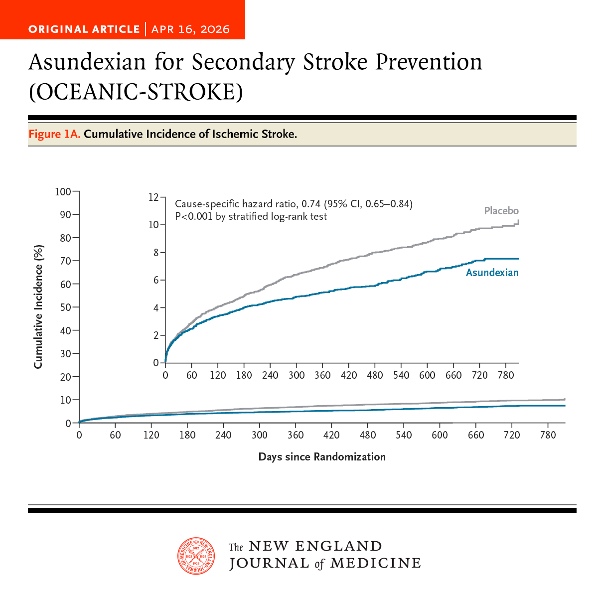
In the phase 3 OCEANIC-STROKE trial involving patients with noncardioembolic ischemic stroke or high-risk TIA, asundexian added to antiplatelet therapy led to a lower risk of ischemic stroke without increasing major bleeding. Full trial results: https://t.co/UPJGedYXFe
Editorial: Asundexian for Noncardioembolic Ischemic Stroke https://t.co/V1dVNbx4T8
Proof Of Concept! 🔥
Yesterday @Tesslagra had an idea, and today we put it to the test!
Pt is a 78 year old with prior CABG and a known wide aortic root. The referring MD had difficulty engaging the LM.
This is our attempt using the hybrid JL/EBU guide! Worked like a charm.😀
The TEMPO-2 trial led to controversy regarding the utility of thrombolysis in patients with NIHSS 0-5 who have an intracranial vessel occlusion. Below is a thread dissecting the trial and available data on minor stroke as well as my interpretation of the trial and practice. 🧵
1/3 Pourfour du Petit Syndrome (reverse Horner syndrome). Is a rare sympathetic nervous system disorder: Ipsilateral mydriasis, upper eyelid retraction and hyperhidrosis.
Dissection of the CCA, during stenting of the ICA. Treated with a #CGuard-stent. Recovered 'ad integrum'