VTE anticoagulation dosing by renal function, from ESC
Apixaban 10mg BID→5mg BID (works even in dialysis at reduced dose)
Rivaroxaban 15mg BID→20mg QD (eGFR≥30 only)
Edoxaban: LMWH lead-in, then 30 or 60mg QD by eGFR
Dabigatran: needs eGFR≥30
🫀 This paper is quietly dangerous. Not because it’s wrong— but because it can be misunderstood.
Let’s go straight to the core.
What the study actually shows
In 479 CCS patients undergoing CCTA:
👉 Lower LDL-C (<70 mg/dL)
was associated with
👉 MORE severe CAD and higher plaque burden
Even more interesting:
👉 These patients had
- more diabetes / pre-diabetes
- more metabolic syndrome
- worse glucose metabolism markers
And:
👉 Diabetes (OR ~6.1) and pre-diabetes remained strong independent predictors of CAD risk
The uncomfortable observation
👉 The “best treated” patients (low LDL) had the worst arteries
Before anyone panics:
This is NOT saying LDL reduction is harmful.
This is saying something much more subtle—and more important.
What is really happening?
1. Reverse causality (the elephant in the room)
Patients with:
👉 prior events
👉 higher baseline risk
👉 more aggressive treatment
→ end up with lower LDL
So:
👉 Low LDL is a marker of treated high-risk patients
Not the cause of disease.
2. Metabolic risk is the real signal
The study shows very clearly:
👉 Glucose dysregulation dominates residual risk
- Diabetes
- Pre-diabetes
- Insulin resistance
- Metabolic syndrome
→ strongly associated with plaque burden and Leiden score
This is the key shift
For years, we simplified CAD risk to:
👉 LDL = bad → lower is better
That’s still true.
But incomplete.
What this paper actually teaches
👉 You can have “perfect LDL” and still have high atherosclerotic risk
Because:
👉 Atherosclerosis ≠ cholesterol alone
👉 It is a metabolic + inflammatory + vascular disease
The imaging angle (the real gold here)
CCTA shows:
👉 more non-calcified plaques in low LDL group
👉 higher plaque burden (SIS)
👉 higher Leiden risk scores
This is exactly where imaging becomes decisive:
👉 Biology > numbers
Clinical implication (the part guidelines are still catching up with)
Treating LDL alone:
❌ does NOT eliminate risk
Because:
👉 residual risk = metabolic + inflammatory + phenotypic
My take
This paper reinforces a concept that is still underused clinically:
👉 CAD is an atheroma disease, not a cholesterol disease
The real mistake to avoid
A superficial reading would lead to:
❌ “Low LDL is associated with more disease → LDL doesn’t matter”
That would be wrong.
The correct interpretation
👉 LDL lowering works
👉 BUT it does not address the whole disease
Where this goes next
This is exactly where:
👉 advanced CCTA (plaque quantification, phenotype)
👉 PCCT (microstructure, composition)
👉 AI-QCT
will redefine risk stratification.
Bottom line
👉 LDL reduction is necessary
👉 but absolutely not sufficient
And if you only track LDL:
👉 you are managing a number
👉 not the disease
I wouldn’t necessarily recommend starting all the cardiology reading before fellowship unless you have extra time during your third year of residency.
That said, the following preparation was extremely helpful for me:
1. Marino's The ICU Book before the CCU rotation.
Cardiac Intensive Care By Dr Brown
A lot of cardiologists gradually lose touch with the intensive care aspect over time. As a fellow, being strong in ICU medicine can really help.
2. EKG books — read as many as you can. My favorites were:
◦The ECG Criteria Book Okeefe
◦Rapid Interpretation of EKG's Dubin
◦The Only EKG Book You'll Ever Need Malcom Thaler
3.I loved using a pocket cardiology book during my first year. I kept adding notes to it, and by third year it had become three times bigger. I did not use it as much later on, but it was invaluable early in training.
4. For echo, buy textbooks by Jae K. Oh, Catherine Otto, and board review by Allan L. Klein. I started reading them from the beginning of fellowship along with the Mayo echo videos, and it helped tremendously.
5. Finally, going home and reading Braunwald's Heart Disease on weekends and late evenings really helped reinforce everything I learned during the day. It is an excellent resource.
@CardioNerds has some excellent resources- wasn’t available when I was a fellow but I hear it’s incredibly useful
Pace yourself. Cardiology is a marathon, not a sprint.
If you are in your third year of internal medicine residency, I would start with the EKG books first.
#Cardiology #FIT #Cardiotwitter #CardioX
1/14
Why can't you use direct oral anticoagulants (DOACs) in patients with mechanical valves (MVs)?
DOACs have been one of the most important advances in my career. And yet, the presence of a MV is one of the few contraindications.
The reason highlights the unique nature of thrombus formation in those with a MV and provides insights into the evolution of human hemostasis.
Coronary Artery Dominance: A Key Concept in Angiography
Dominance is defined by which artery gives rise to the posterior descending artery (PDA):
• Right dominance – PDA from RCA
• Left dominance – PDA from LCx
• Balanced – PDA from both RCA & LCx
Prevalence:
• Right dominant – ~85%
• Left dominant – ~8%
• Balanced – ~7%
Understanding dominance is crucial in assessing myocardial perfusion, planning revascularization, and anticipating outcomes in coronary artery disease.
Image:
Top – Right dominance
Middle – Left dominance
Bottom – Balanced dominance
(Courtesy Dr. Annapoorna Kini, Mount Sinai, NY)
Ref: Braunwald’s Heart Disease, 12th Ed.
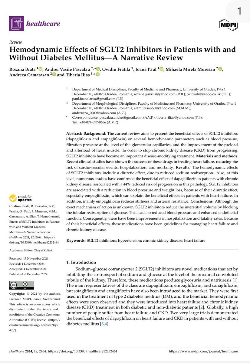
❤️ SGLT2 inhibitors are not “diabetes drugs” anymore...They are hemodynamic drugs!!
We’ve all seen the shift in guidelines.
But many still think:
👉 “They lower glucose → good for diabetics” 🚨 That’s outdated thinking. 📚 A recent narrative review clearly reinforces what we’re already seeing in practice:
⚙️ SGLT2 inhibitors fundamentally reshape hemodynamics
🧪 At the kidney level:
▪️ ↓ Sodium reabsorption
▪️ ↑ Natriuresis + osmotic diuresis
▪️ ↓ Glomerular pressure (via tubuloglomerular feedback)
👉 Translation:
✔️ Less hyperfiltration
✔️ Slower CKD progression
✔️ Better renal protection
❤️ At the cardiovascular level:
💧 Preload reduction
👉 Interstitial fluid loss > intravascular depletion
✔️ Decongestion without aggressive hypotension
🫀 Afterload reduction
👉 ↓ Arterial stiffness
👉 ↓ systemic vascular resistance
📉 Blood pressure effect
👉 Modest but consistent (~3–4 mmHg systolic)
✔️ Without reflex tachycardia
✔️ Without RAAS overactivation
🔥 The real magic:
👉 Unlike loop diuretics:
🟥 Loop diuretics → intravascular depletion → neurohormonal activation
🟩 SGLT2 inhibitors → interstitial unloading → hemodynamic stability
📊 Clinical outcomes back it up
✔️ ↓ HF hospitalization
✔️ ↓ CV mortality
✔️ ↓ CKD progression (~44% in some cohorts)
🎯 The paradigm shift
These drugs are:
❌ Not just antidiabetics
❌ Not just mild diuretics
✔️ They are cardio-renal modulators
💡 Clinical takeaway
👉 Think SGLT2 inhibitors when you think:
▪️ Heart failure (HFrEF + HFpEF)
▪️ CKD (with or without diabetes)
▪️ Cardio-renal syndrome
🧠 And most importantly:
👉 Use them early
👉 Not as “last-line add-ons”
🔥 The future of heart failure is not just inotropes and diuretics.
It’s metabolic-hemodynamic modulation.
💬 Your experience:
👉 Are you already using SGLT2 inhibitors systematically in non-diabetic HF patients?
👉 Or still waiting for “perfect indications”?
📚 Reference
Brata, R., etc al. Healthcare, 12(23), 2464. https://t.co/i7zVeyOM0B
“Wellens” is one of the most overcalled ECG patterns on #FOAMed. Sometimes there is also incidental bystander stenosis on the angiogram.
A simple check: does the ECG change significantly after PCI? If not, it’s far more likely TWI is LVH/HCM or structural.
#STEMI
61 y old hypertensive lady with chronic AF presented with acute chest pain at 3 am .
#ECG inferior STEMI
Coronary angiogram 👇
Stent or not ?
#cardiology#CardioEd
Case for fellows. Patient admitted with recent onset heart failure. She was detected to have a murmur 3 years ago, but has been asymptomatic till 2 weeks back. Your observations?
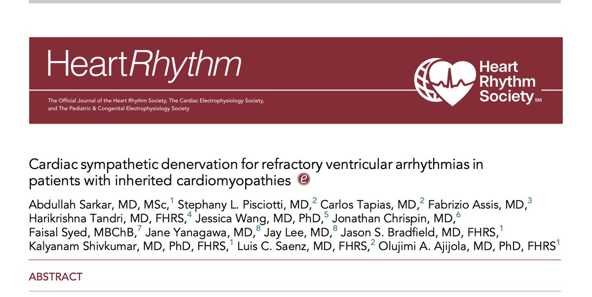
⚡ When drugs and ablation fail… target the nervous system
We are used to thinking about ventricular arrhythmias as:
👉 scar
👉 fibrosis
👉 re-entry
But this paper reminds us of something fundamental:
👉 arrhythmias are also neuro-driven
🧠 The concept:
If sympathetic activation triggers arrhythmias…
👉 why not remove the trigger?
➡️ Cardiac sympathetic denervation (CSD)
📊 In patients with inherited cardiomyopathies (HCM, ACM, DCM):
• Highly refractory population
• Failed drugs + multiple ablations
AND YET 👇
✔️ ICD shocks ↓ 61%
✔️ ~50% arrhythmia-free
✔️ 76% transplant-free survival at 4 years
1-s2.0-S1547527125030371-main
🔥 Look at the figure (page 4):
👉 Almost every patient had fewer shocks after CSD
Not zero.
But meaningfully less
⚡ This is the real value:
Not cure.
👉 modulation
🧬 Why does it work?
Because sympathetic tone:
• lowers arrhythmia threshold
• increases triggered activity
• destabilizes substrate
👉 CSD removes that adrenergic drive
⚠️ But here’s the key nuance:
❌ It doesn’t fix the disease
❌ It doesn’t remove scar
👉 It changes the environment where arrhythmias occur
💥 And one important insight from the paper:
👉 Heart failure — not arrhythmia —
is still the main driver of outcomes
🎯 Clinical take-home
When you face:
• recurrent ICD shocks
• VT storm
• failed ablation
👉 Think beyond myocardium
👉 Think autonomic system
💬 Are we underusing neuromodulation in arrhythmia management?
#Cardiology #Arrhythmia #VT
#Cardiomyopathy #Neurocardiology
#EPeeps #CardioTwitter ⚡🫀
https://t.co/MJFqJRSZ6z
Among patients with anterior myocardial infarction, adding low-dose rivaroxaban to dual antiplatelet therapy did not significantly reduce left ventricular thrombus formation at 1 month but increased minor bleeding.
https://t.co/TDDR0p8sWV
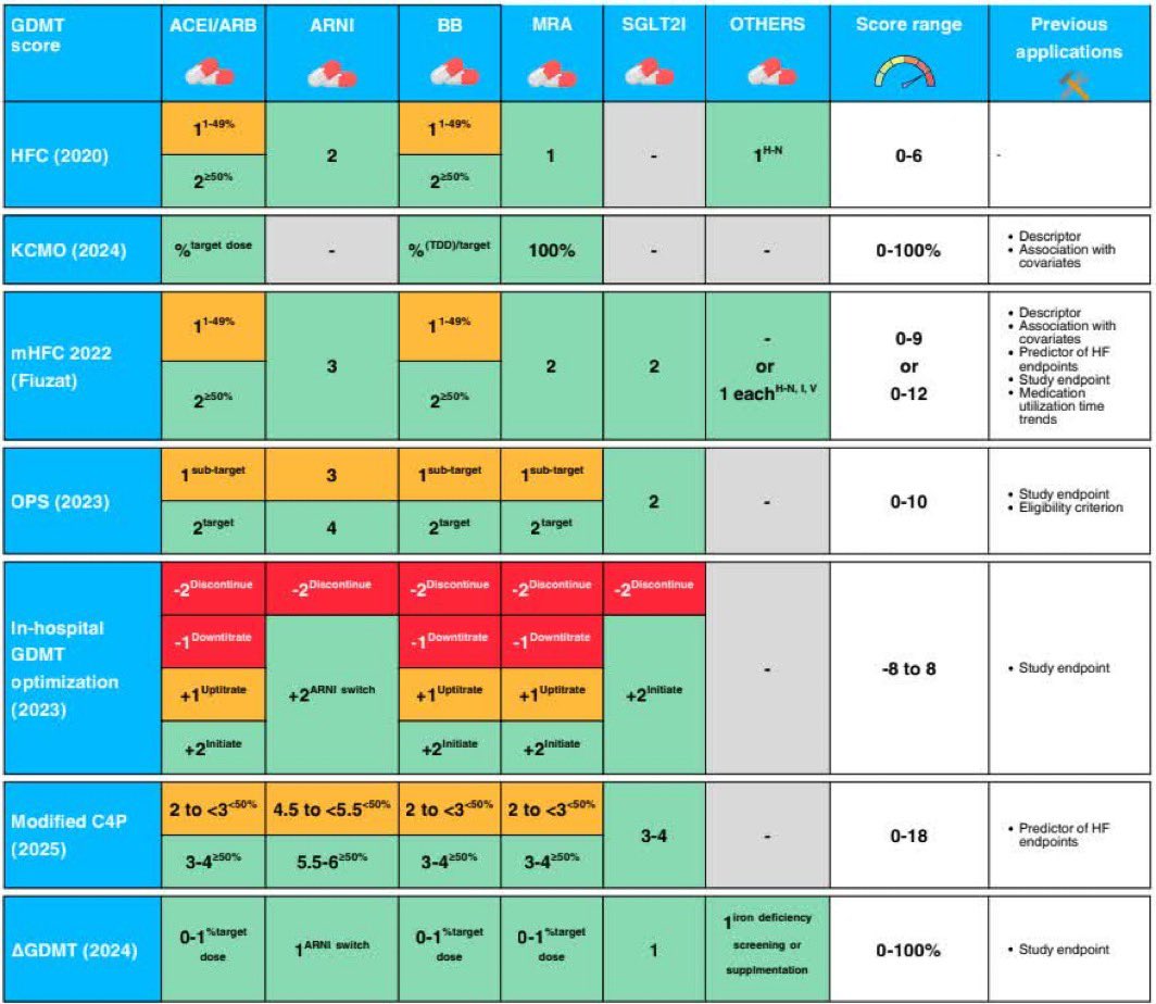
Heart Failure Guideline-Directed Medical Therapy Scoring Systems: A Scoping Review
Multiple, disparate scores have been developed to quantify heart failure with reduced ejection fraction GDMT optimization, undermining the original intent. Key gaps in scores include heterogeneous weighting of drug classes and doses, minimal incorporation of intolerance to identify maximum-tolerated therapy, and heterogeneous incorporation of therapies beyond quadruple therapy
#Cardiology #MedTwitter #CardioTwitter #HeartHealth #Healthcare
@mvaduganathan@SJGreene_md@DrMarthaGulati@hvanspall@gcfmd@hfcollaboratory@ShahzebKhanMD@safchat
https://t.co/31XLTszj3A
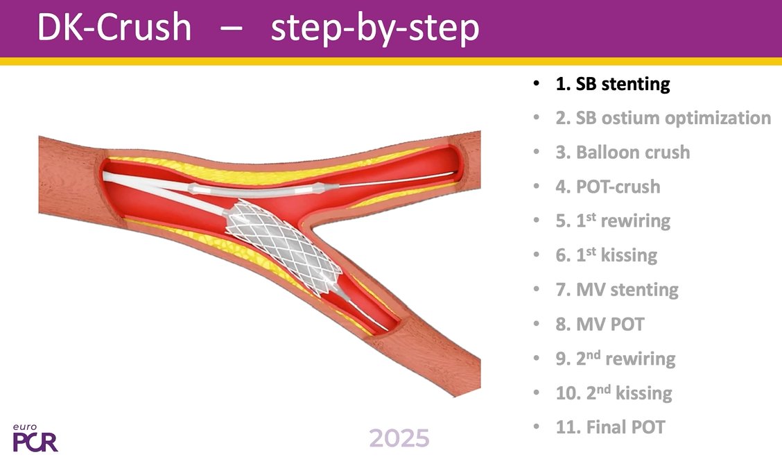
📢DK Crush like you’ve never seen it before!
Episode 1⃣ of this series on "How to perform DK-Crush for left main bifurcation stenting" now online
📺https://t.co/Y64RCYU1w3
In this episode, learn about:
🟣Multi-modality simulation to unveil the hidden parts & small obstacles that can result in complications
🟣Angioscopy directly inside the coronary artery to provide a better understanding of 3D stent configuration
In-depth commentary and perspective and perspective provided by @GoranEBC & William Wijns.
@twj1974@NievesGonzalo1@BURZOTTA_F@M_Lesiak@gabor_gt@RhianEDavies1@esbrilakis@VisibleHeartLab
#interventionalcardiology #CardioEd #EuroPCR