A great week of science (and gelato🍦) in Padua! 🇮🇹
Grateful for the opportunity to present part of my PhD research at #IGJC2026 and share my work with the gap junction community.
Lots of great conversations, new connections, and ideas to bring home!
Park et al. distinguish two subtypes of dementia with Lewy bodies based on distinct imaging biomarker trajectories. One is characterised by early striatal dopamine loss and more typical Lewy body features, while the other shows early amyloid accumulation. https://t.co/g1qVFX0ga0
Grateful to have presented my research at the 4th Canadian Zebrafish Research Community Meeting and honoured to receive the Best Oral Presentation Award!
Thank you to the organizers for a great meeting, and congratulations to all presenters!
Neuroimmune dysregulation in Alzheimer's disease: Mechanisms and therapeutic strategies
"The perspective shifts from traditional Aβ/tau models to recognize neuroimmune dysregulation as a core mechanism of AD."
https://t.co/2lywEmVXqb
Most people who take CoQ10 think of it as an antioxidant. It is one. But that is not the most important thing it does.
CoQ10 is the only mobile electron carrier in the inner mitochondrial membrane. The electron transport chain has four protein complexes fixed in the membrane. Complex I accepts electrons from NADH. Complex II accepts them from FADH2. But neither can pass those electrons directly to Complex III. They hand them to CoQ10, which physically shuttles across the lipid bilayer to deliver them. Complex III passes them to Complex IV, which reduces oxygen to water and drives the proton gradient that ATP synthase uses to produce ATP.
Without CoQ10, the chain breaks between Complex I/II and Complex III. Electrons have nowhere to go. The proton gradient collapses. ATP production stalls. This is not an antioxidant function. This is the core mechanism of aerobic energy production.
CoQ10 is predominantly synthesized endogenously through the mevalonate pathway, the same pathway that produces cholesterol. HMG-CoA reductase is the rate-limiting enzyme. Statins inhibit HMG-CoA reductase. That is how they lower cholesterol. It is also how they lower CoQ10.
An updated meta-analysis by Qu et al. (2018) pooled 12 RCTs with 1,776 participants and found statins significantly reduced circulating CoQ10. The reduction was independent of statin type, intensity, or treatment duration. Both lipophilic and hydrophilic statins produced the same effect. This is consistent with what the biochemistry predicts: the pathway is shared.
On top of statin-induced depletion, CoQ10 in human heart tissue declines naturally with age. Kalén et al. (1989) measured CoQ10 concentrations in myocardial tissue and found levels peak around age 20, decline by more than 30% by age 40, and drop approximately 50% by age 80. The organ with the highest energy demand loses half its electron carrier over a lifetime.
A 2025 meta-analysis by Kovacic et al. (Journal of Nutritional Science, 7 RCTs, 389 patients) found CoQ10 supplementation significantly reduced statin-associated muscle symptoms measured by pain intensity. This is the most current pooled data on clinical outcomes.
One important nuance: while plasma CoQ10 depletion from statins is well established, whether intramuscular CoQ10 drops proportionally is inconsistent. Some studies found no change or even increases in muscle tissue CoQ10 during statin treatment. The plasma reduction may partly reflect reduced LDL particles, which are the primary carriers of CoQ10 in blood. The clinical significance of depletion beyond muscle symptoms remains debated.
Roughly 200 million people worldwide take statins. The mevalonate pathway that produces their target also produces the electron carrier their mitochondria depend on. The mechanism is not controversial. The clinical implications are still being defined.
Kalén et al., Lipids, 1989.
Qu et al., Eur J Med Res, 2018.
Kovacic et al., J Nutr Sci, 2025.
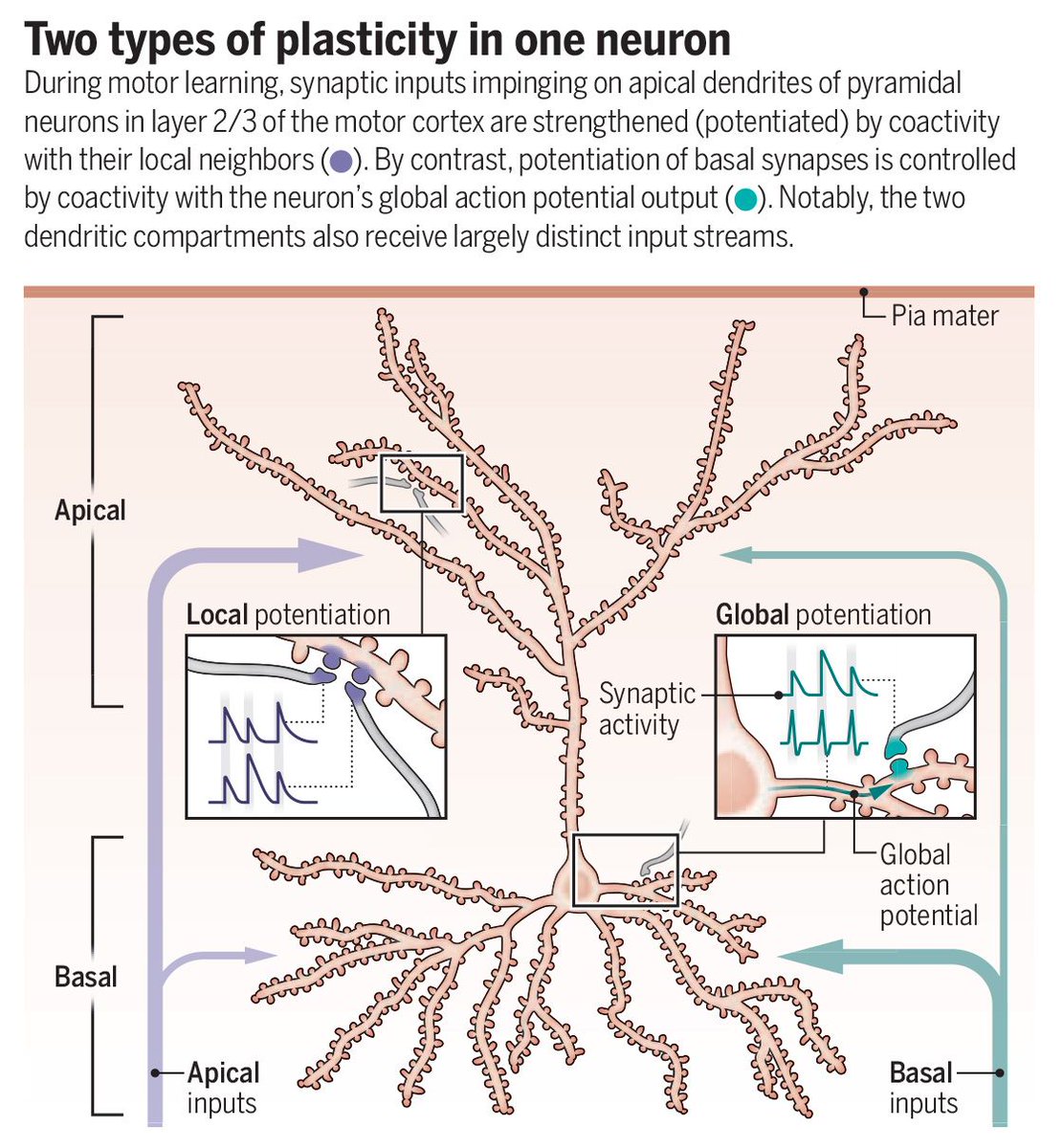
A 2025 Science study found that different dendritic segments of a single neuron follow distinct rules.
The results challenge the idea that neurons follow a single learning strategy and offer a new perspective on how the brain learns and adapts behavior
Learn more in this #SciencePerspective: https://t.co/FwMKmSnYBC #ScienceMagArchives
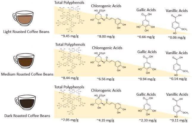
Coffee roasting changes the content of chlorogenic acids and other antioxidants
Coffee beans are packed with chlorogenic acids - plant compounds that help regulate blood sugar, protect blood vessels, and act as antioxidants. Roasting changes how much of these (and other polyphenols) end up in your cup:
1️⃣ Light Roast
Highest total polyphenols (~9.45 mg/g).
Chlorogenic acids dominate (~8.0 mg/g).
🟢 Example: Lighter roasts keep the most chlorogenic acids, which may support glucose control and vascular health.
2️⃣ Medium Roast
Slightly fewer polyphenols (~8.44 mg/g).
Chlorogenic acids fall (~6.56 mg/g), while gallic acids rise (~0.94 mg/g).
🟢 Example: Some chlorogenic acids break down, but roasting creates new antioxidant compounds like gallic acid.
3️⃣ Dark Roast
Lowest total polyphenols (~7.95 mg/g).
Chlorogenic acids drop sharply (~4.35 mg/g).
Gallic acids peak (~2.33 mg/g).
🟢 Example: Dark roasts lose much of their chlorogenic acid but swap in different antioxidant chemistry from roasting.
Chlorogenic acids = the main health-promoting polyphenols in coffee, highest in light roast.
Gallic acids increase with darker roasts, bringing a different antioxidant profile.
The roast you choose shifts the balance, but all deliver unique benefits
Top-down selection of visual working memory contents is supported by alpha-band phase-synchronized oscillatory networks https://t.co/2sXtAUiHRe
#neuroscience
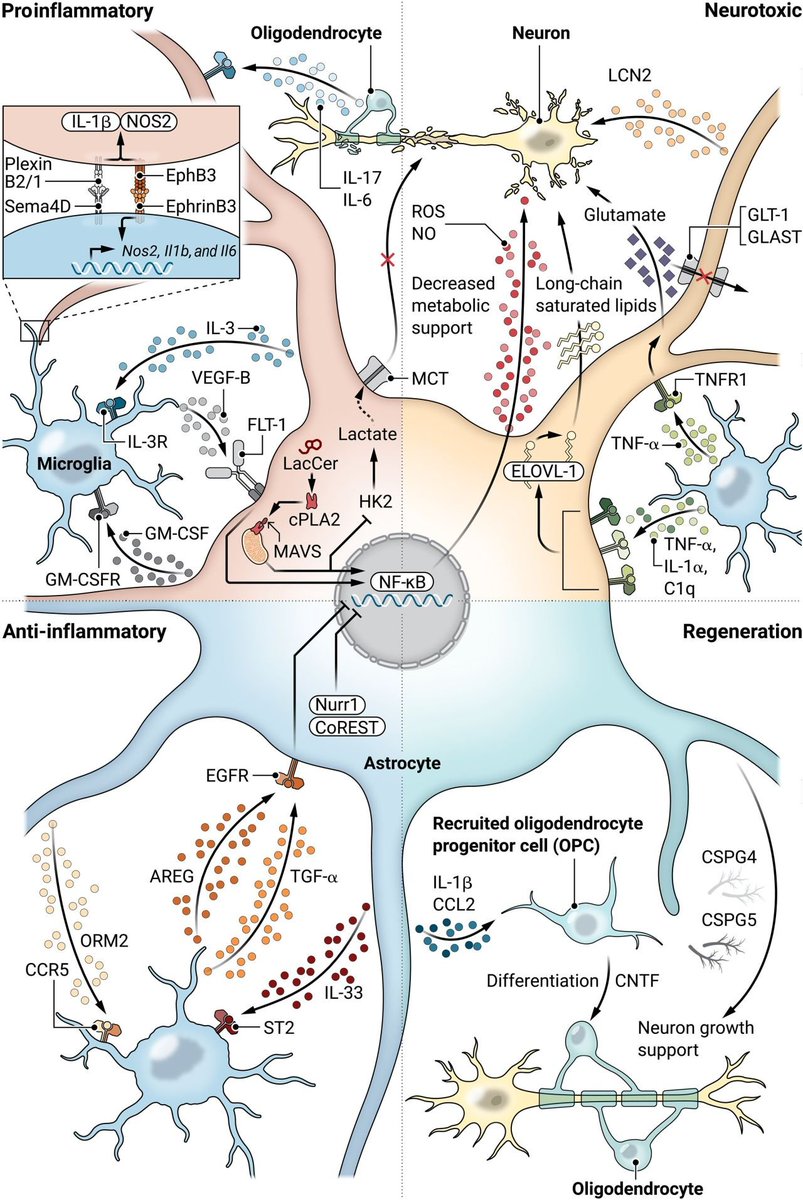
Our brain isn’t just made of neurons. It runs on a whole neighborhood of cells that can either protect your mind or quietly push it toward inflammation.
This diagram reveals how astrocytes, microglia, neurons and oligodendrocytes talk to each other during stress, illness, and injury. Their conversations shape memory, mood, cognition, and long-term brain resilience.
Here is what this graphic shows in plain language:
🧠 Astrocytes act as the central switchboard
They decide whether the brain environment becomes supportive or inflammatory. When they sense danger signals, they activate NF kappa B and release molecules that influence the other cells around them. When conditions are safe, they release factors that help neurons grow and help new oligodendrocytes mature.
🔥 Microglia can protect or damage depending on the signals they receive
They can release IL 1 beta, GM CSF, and other proinflammatory signals, or they can shift to a more supportive state depending on what they detect from astrocytes and neurons.
⚡ Neurons suffer when inflammation rises
Oxidative stress, nitric oxide, and loss of metabolic support weaken them. Glutamate handling becomes impaired, which increases excitotoxic stress.
🧩 Oligodendrocytes and their precursor cells respond to what the environment tells them
Inflammatory signals slow their support for neurons, while regenerative signals encourage new myelin formation and better neuron stability.
The health of your brain depends on how these cells interact, not just on neurons alone.
doi: 10.1126/scitranslmed.adi7828
Although new AD therapies reduce amyloid plaques, they have not led to major clinical improvements. Pini et al. suggest that changes in brain connectivity may offer a more sensitive and biologically meaningful marker of disease modification. https://t.co/jVpaiXz4zI
Our brain isn’t just made of neurons. It runs on a whole neighborhood of cells that can either protect your mind or quietly push it toward inflammation.
This diagram reveals how astrocytes, microglia, neurons and oligodendrocytes talk to each other during stress, illness, and injury. Their conversations shape memory, mood, cognition, and long-term brain resilience.
Here is what this graphic shows in plain language:
🧠 Astrocytes act as the central switchboard
They decide whether the brain environment becomes supportive or inflammatory. When they sense danger signals, they activate NF kappa B and release molecules that influence the other cells around them. When conditions are safe, they release factors that help neurons grow and help new oligodendrocytes mature.
🔥 Microglia can protect or damage depending on the signals they receive
They can release IL 1 beta, GM CSF, and other proinflammatory signals, or they can shift to a more supportive state depending on what they detect from astrocytes and neurons.
⚡ Neurons suffer when inflammation rises
Oxidative stress, nitric oxide, and loss of metabolic support weaken them. Glutamate handling becomes impaired, which increases excitotoxic stress.
🧩 Oligodendrocytes and their precursor cells respond to what the environment tells them
Inflammatory signals slow their support for neurons, while regenerative signals encourage new myelin formation and better neuron stability.
The health of your brain depends on how these cells interact, not just on neurons alone.
doi: 10.1126/scitranslmed.adi7828
#ZebrafishFunFacts: Known to hobbyists as the leopard Danio, TL or Tüpfel longfin #zebrafish consist of two alleles (leo and lof). leo is a gap junction protein (https://t.co/SedSLCVqKp), while lof is caused by upregulation of a K+ channel (https://t.co/RydgQu43NJ). 1/2
Three conferences and a Symposium this year! Grateful for the chance to share my research at the 3rd CZRC, CAN 2025, 50th AGSBS, and now at the CVR-CIAN conference hosted at York University.