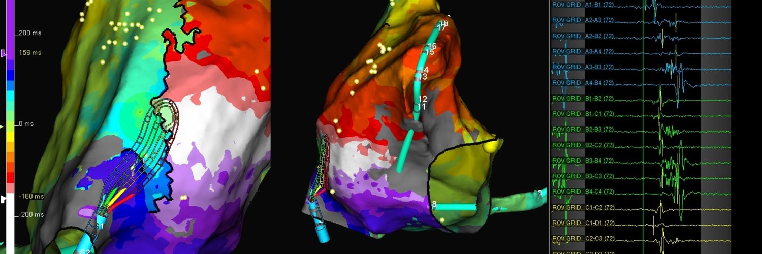
First case using #GridX with the 2 new sensors on the tip. Mapped so much faster than regular Grid, due to it being in high confidence 100% of the time, with way less geometry editing. But the best thing is no more having to say "We need more voxels"! 😂 #EPeeps
With multi-wavefront mapping in VT ablation we are always looking for new ways to visualize core concepts. Emphasis mapping on the activation window with ILAM highlights the narrowest isochrones to visualize WADLs which co-localize with the 3D boundaries of the VT. (Thread 1/6).
HD Grid X making her worldwide debut at #VTSymposium. Two additional SE sensors on the distal portion of the Grid allow for faster and more accurate modeling and mapping immediately upon insertion.
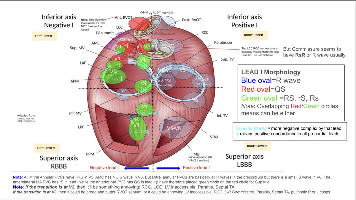
PVC localization diagram. Added my own touches. adapted from the nice figure by Enriquez et al. Illustrates the precordial transitions to positive complexes, lead I morphology (blue/red/green circles), and precordial transition to negative complexes. #EPeeps
NEW ICE Image of the Month: To highlight AFib Awareness Month, #HRStv host @DanielAlyeshmer is joined by @Drdevignair to discuss the novel application of ICE for PFA ablation of AFib. They discuss ICE imaging for PFA ablation for PVI and extrapulmonary triggers. See the full interview here #EPeeps ➡️ https://t.co/aSbEEssc2j
Correlation between VT isthmus, imaging findings and histopathology in a patient with desmoplakin cardiomyopathy.
A collaborative report with @ColumbiaPS pathology team published in @JACCJournals#JACCEP@WeillCornell
https://t.co/WsUSwL8j8r
Here is a video clip of Papillary muscle PVC ablation showing how #ICE provides real-time visualization of cardiac structures and continuous monitoring of catheter location. Please turn the sound on.
We have had a great initial experience with Farapulse and Ensite X! The mapping system coupled with ICE in the left atrium ensures proper catheter positioning for therapy delivery. @AbbottCardio@Segarspr@BSCCardiology @WilliamMaddoxMD
Post-myocarditis #VT in a young patient with recurrent syncope.
👇
ILAM demonstrating color crowding in both sides of narrow corridor corresponding to entrance & exit of clinical VT.
#EPeeps
The value of ICE in epicardial VT ablation.
1. Ensure epicardium dry before ablating to ensure optimal lesion delivery.
2. Understand scar and substrate in three dimensions.
3. Monitor for complications during access (visualize wire course) and throughout case (bleeding).
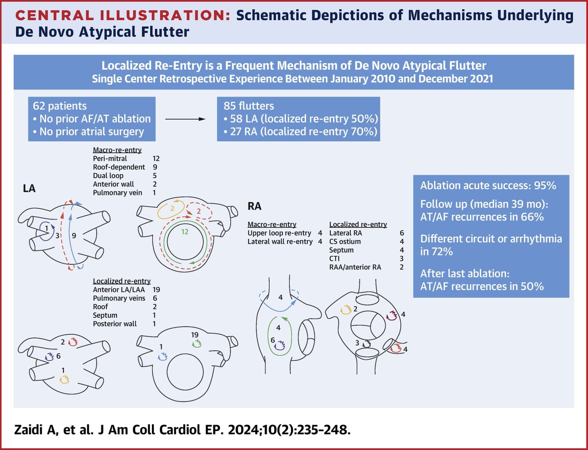
Atypical atrial flutters in pts w/out prior surgery or complex ablation are often due to localized re-entry. Other ATs commonly occur during long-term follow-up following ablation, suggesting progressive atrial myopathy in these pts. https://t.co/yknvY85Jkm
#JACCCEP#epAblation
Remarkable agreement between ILAM deceleration zone, CT wall thickness channel, and peak frequency emphasis on the apical scar. Case by @davilandre and Dr. Bahij Kreidieh. Ensite mapping by @DavisSneider with @ADAS3D integration. #AblateVT
“Peak Frequency” annotation based voltage map using Near Field OT helps to identify the sites critical to #VT circuits.
👇
Endocardial map of non-ischemic VT after MV repair demonstrating low voltage-high frequency sites colocalized with critical isthmus of VT. #EPeeps#ablateVT
Elegant work on the analysis of EGM morphology. The conventional use of a "balanced" EGM signal does not identify the anatomical mitral valve annulus. The A/V EGM ratio between 1:9 and 1:16 best approximates the mitral valve annular plane. Nice work https://t.co/iy9BSQoAIy