Very excited to have our work on spatial profiling of lung precursor lesions and invasive lung cancers out in @Cancer_Cell. This work was done in joint collaboration with @IamLinghua and closely with many esteemed groups. Here is a run down of our major findings!
We studied multiple cohorts of normal lung tissues, precursor/preinvasive lesions and invasive lung cancer by multiple single-cell and spatial profiling modalities. We first find that invasive lung cancers, naturally, have more complex spatial expression patterns than their premalignant counterparts.
Phylogenetic reconstruction using our spatial transcriptomics analysis found that the earliest clones mapped to reactive pneumocytes, the histological resemblant of KRT8+ alveolar cells (KACs)/intermediates! This was super exciting! We previously reported that these transitional cells were not so transitional, they were stuck in limbo and acted as progenitors of lung adenocarcinoma.
KACs displayed spatial metaprograms that were distinct from normal alveolar cells and that were closely related to premalignant lesions. Remarkably, KACs had uniquely high expression of 'drivers' of inflammation (IL1R1) and were present in niches that were rich with IL1B high myeloid (macrophages) cells.
This epithelial-proinflammatory niche was disease stage-specific. Unlike oncogenic properties which we believe accumulate over the lifetime of the lesion, these niches peaked in early precursor lesions and faded in more advanced stages.
We functionally validated these KAC/epithelial-proinflammatory niches in lung carcinogenesis models showing that IL-1b treatment or co-culture with interstitial macrophages act as mitogens for KRT8+ high alveolar cells.
Importantly, there is preclinical value (and certainly clinical) value for our findings. We found that targeting IL-1B by neutralizing antibodies, including when combined with PD-1 blockade, was effective in preventing formation of precancerous lesions and their progression to adenocarcinomas. These effects were associated with reduced abundance of KACs!
Inflammation appears to be operative in the earliest stages of lung adenocarcinoma development and likely drives oncogenesis of alveolar intermediate cells that function in tissue homeostasis/remodeling after injury. Indeed, it is long thought that tumors are wounds that do not heal, and it is in this healing process if chronic where the budding tumor can hijack inherent properties in the lung. It is also plausible that targeting inflammation is valuable for intercepting lung cancer rather than treating the disease in advanced stages. This supposition is supportive of earlier clinical studies with the IL-1B antibody canakinumab (CANTOS).
Very proud of Fuduan Peng joint fellow with the Wang group, now director extraordinaire @Squirrel_PhD, talented student @Yibo_Dai for leading this work, with strong earlier effort from @warapen. I am grateful for collaborations with many groups that I personally learned so much from. This work could not have been done without funding from @NIH@theNCI (NIH funding is important!), @CPRITTexas and @LUNGevity. Also thanks to the astute reviewers as well as the editors @Cancer_Cell who helped us improve our work. #lungcancer #endcancer @MDAndersonNews
https://t.co/YO15Wsrlpc
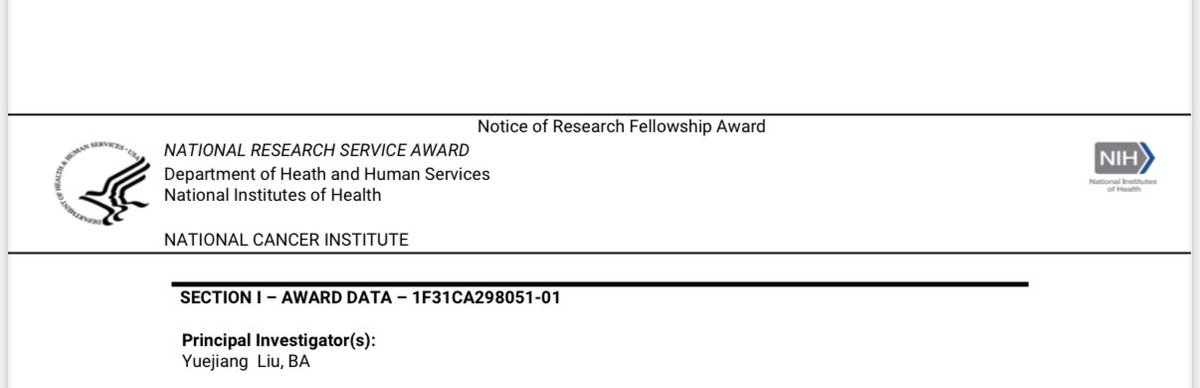
Thrilled to share that I received an NIH F31 grant award! 🥰🥰Huge thanks to my incredible mentors & colleagues at @humam_kadara lab and @MDA_UTHGrad for their unwavering support. Excited to advance my research in dissecting the role of gut microbiome in lung cancer development!
Please check out @ZahraaRahal minisymposium presentation (abstract 6384) at #AACR25 in which she finds a target that mediates development of KRAS-mutant #lungcancer and response to KRAS-targeted therapy
Overwhelmed with gratitude to have matched into Internal Medicine at @BrighamMedRes! This moment belongs to everyone who believed, loved, and lifted me, but most of all to @humam_kadara, my mentor, my compass, and the fiercest believer in me since my premed days. #Match2025
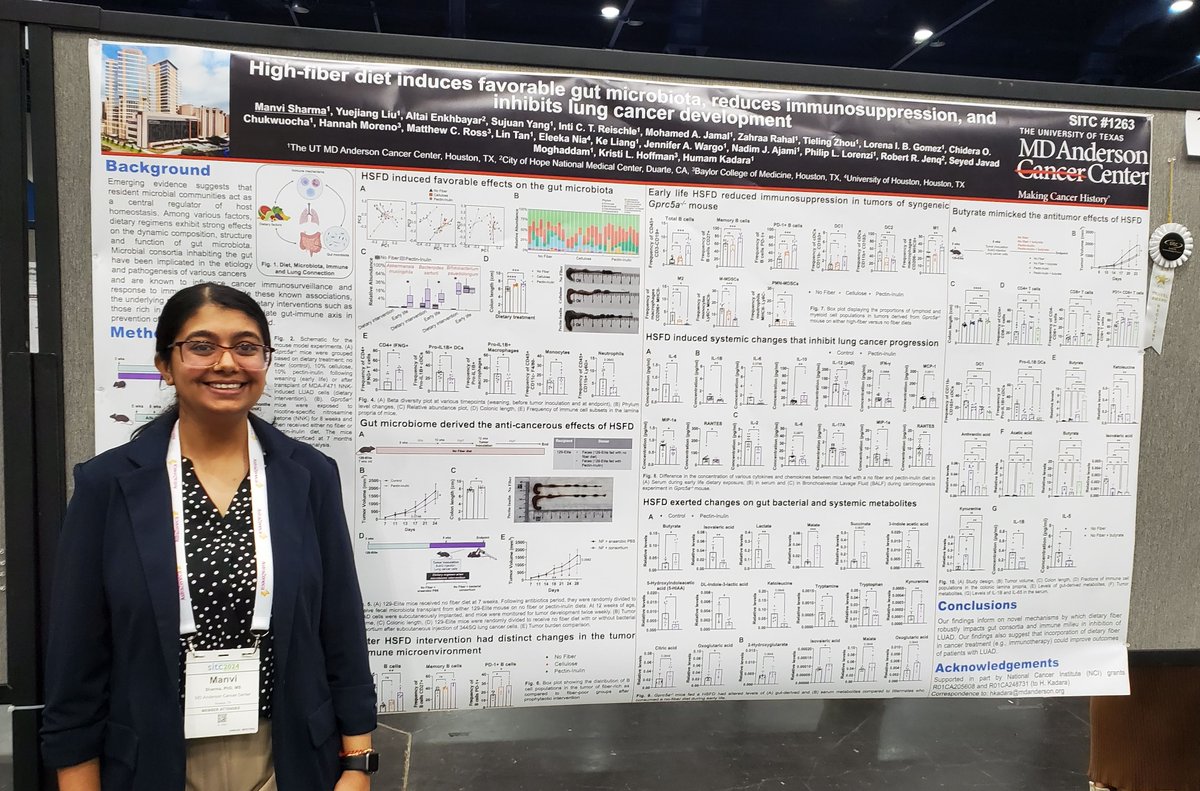
Our postdoctoral fellow Dr. @Manvi_Sharma1 of MD Anderson’s Dr. @Humam_Kadara lab shared findings on the impact of dietary fiber on the gut microbiome, with implications for effects on immunotherapy response (Abstract 1263). @SITCancer#SITC24#EndCancer
Please check our group’s poster 1263 at #SITC24 on effects of high fiber diet on the gut #microbiome, tumor immunity and lung cancer development. Congrats to postdoc fellow in our group @Manvi_sharma1 for presenting this work today and receiving a SITC young investigator award!
Excited to finally share our @nature publication! A thrilling discovery, yes, but for me, a trainee privileged to contribute, it’s a tale of perseverance, teamwork, and personal growth at every conceivable level. Kudos to the @Humam_kadara lab family and collaborators ✨!
Today’s the day - it’s finally out in @Nature! @humam_kadara, I still remember your words in the tissue culture room at the end of a long day & when our data was pointing to us a new discovery: trust the process & don’t give up!
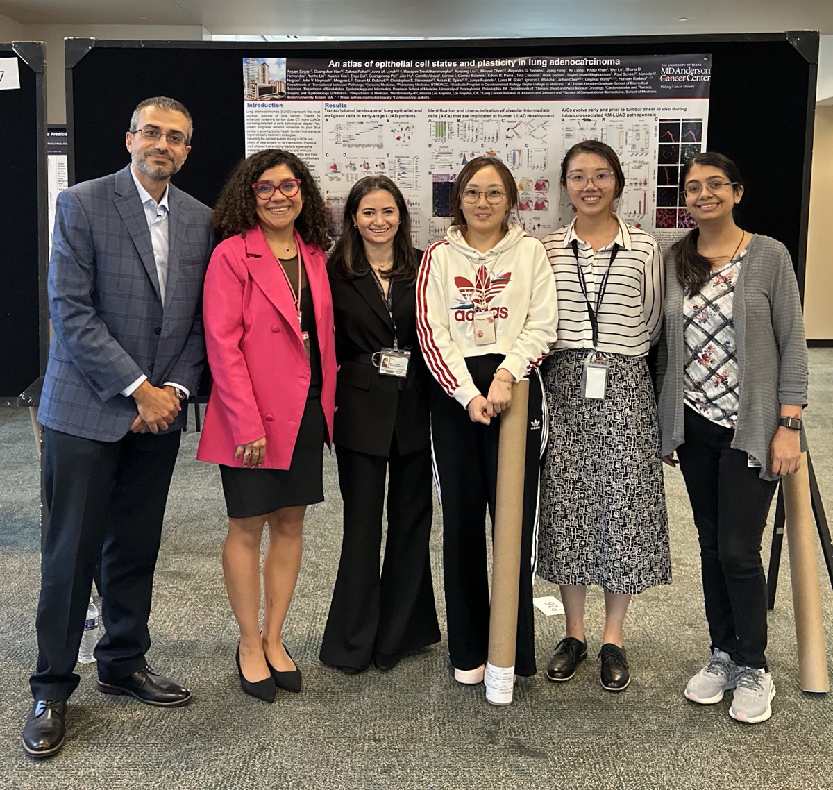
At last, at long last, our labor of love is out today in @Nature! 🥳By generating an atlas of normal and cancerous epithelial cells in lung adenocarcinoma we unearth cellular states/subsets that underlie inception of the disease. https://t.co/fKwADUpaCl. A thread! 1/19
Elated by @Humam_Kadara's lab success at TMP's annual retreat @MDAndersonNews! His Mentorship Award, so rightfully earned, is echoed in us trainees—myself, @Squirrel_PhD, & @Manvi_sharma1—sweeping 1st, 2nd & 3rd in the postdoc category🥇🥈🥉. A true team triumph! #ProudTeamMember