Our resource on the regulation of #RhoGTPase signaling is online. All (!!!) RhoGEFs and RhoGAPs analyzed. I'm sure there is something for everyone in it. Thanks to @rocks_oliver@e_petsalaki @BakalLabICR @PertzL @JulianeRdmchr @RickBagshaw & many more...
https://t.co/E4s9MbRe7M
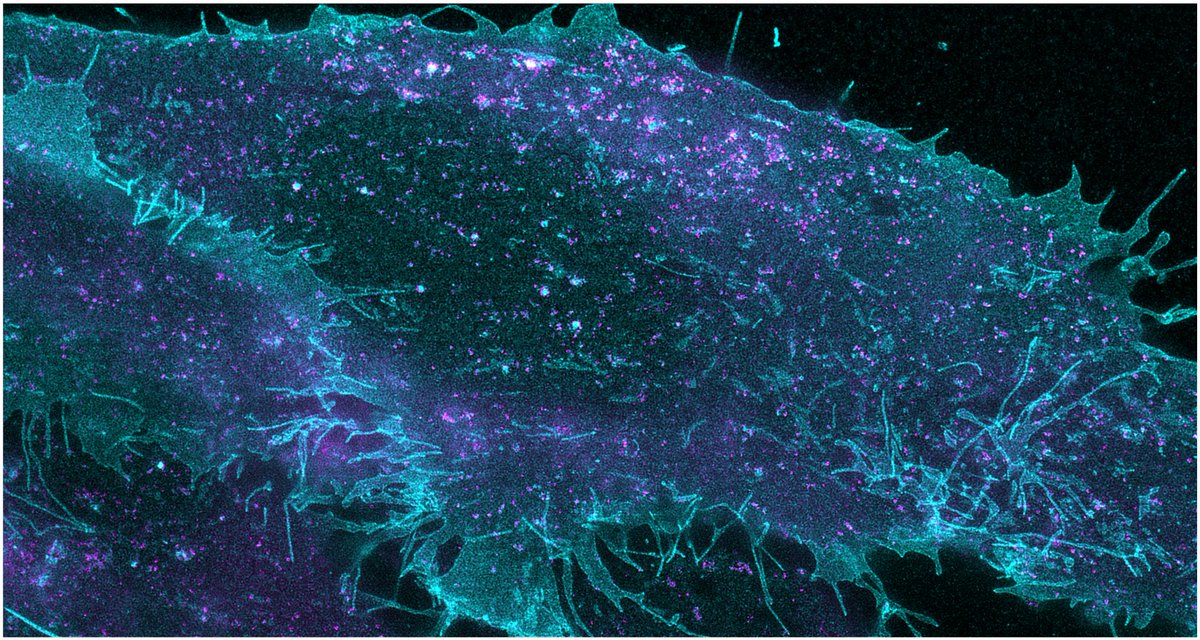
Here’s the big one: In collaboration with @Honigmann_Lab, @LabShevchenko, Björn Drobot and Martin Hof we present a general workflow for imaging the localization and transport of individual lipids in cells and mapping their metabolism.
https://t.co/uotVrHnbsK
Here’s the big one: In collaboration with @Honigmann_Lab, @LabShevchenko, Björn Drobot and Martin Hof we present a general workflow for imaging the localization and transport of individual lipids in cells and mapping their metabolism.
https://t.co/uotVrHnbsK
Working on this with @RiaThielhorn and @ewers_helge fundamentally deepened my understanding of #ExM. I hope it will be useful for others who want to get started in #Expansion#Microscopy 🔬🦠https://t.co/NzJpzZEOHD
Amazing work! @RaluucaG is an impressively talented scientist and it was a great experience to be involved in this project. PIs keep your eyes open for applications.
Glad to see my PhD work from the @ewers_helge lab out now in @NatureComms. Check it out if you want to know more about the biophysics behind lipid-mediated endocytosis.
Glad to see my PhD work from the @ewers_helge lab out now in @NatureComms. Check it out if you want to know more about the biophysics behind lipid-mediated endocytosis.
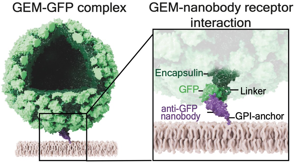
Paper out: Gene encoded nanoparticles (GEMs) with 180 GFPs are cool tools to track in cells. GEMs reveal biophysical basis of infection, too. Using GPI-anchored nanobody receptors over 6 logs of affinity shows that adhesion energy alone explains uptake! https://t.co/cK6dZeOCed
ExM-review online!
@MforMicroscaya @RiaThielhorn and I describe how ExM works, how to do experiments and control them properly. Plus list of everything you need to know and where to find it!
Thx to @AliHShaib Silvio Rizzoli & @HofkensLab for sharing data.
https://t.co/eyzrL1lsg1
Finally it is out 😁. Our review on #expansion microscopy summarizes the key steps of sample prep and gives an overview of the different methods. Perfect for novices to the field 🧑🏽🔬
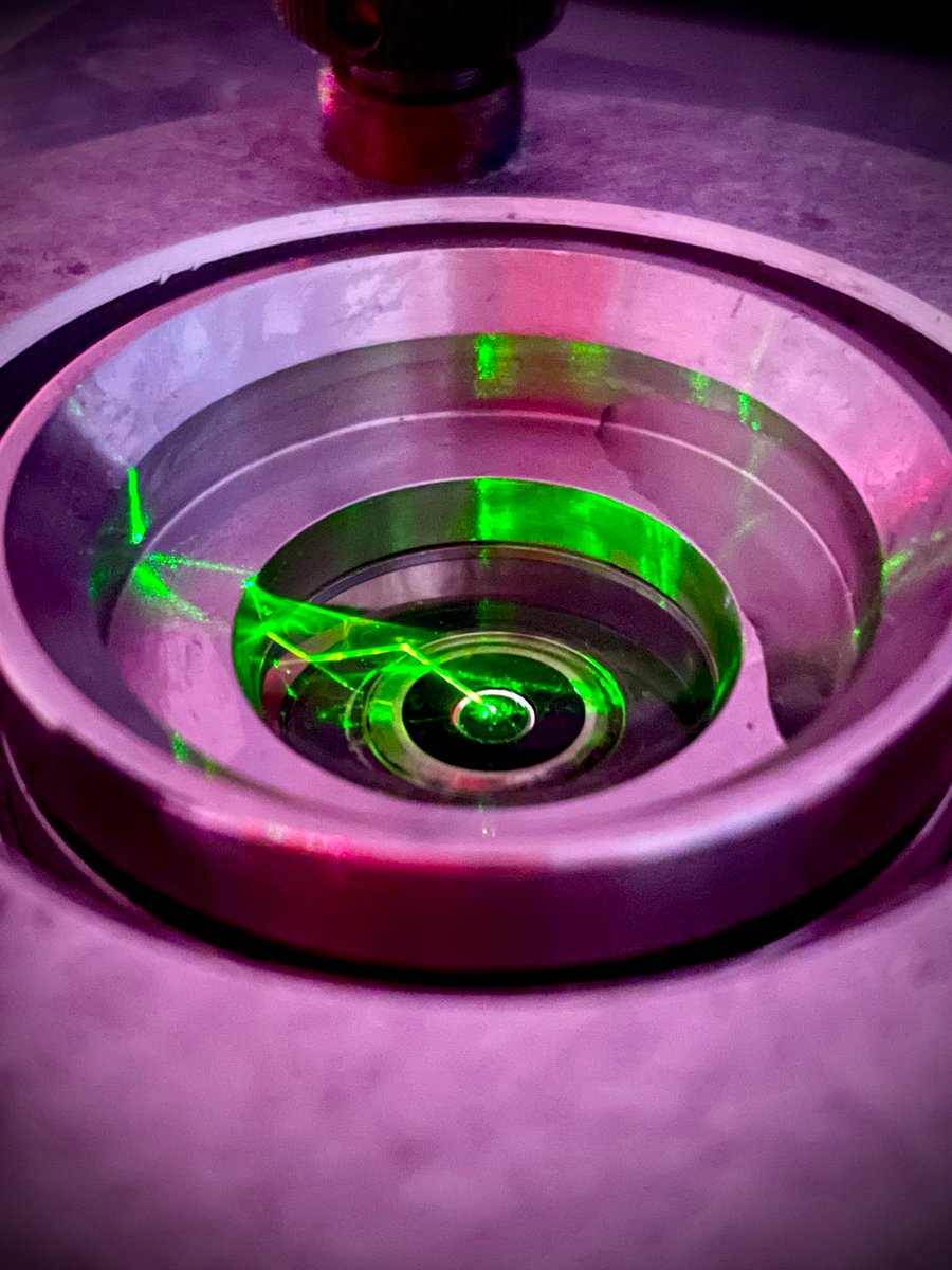
Shine some UV light and a vesicle eats a condensate. Switch it off, and it releases it. Here is how this works (Adv Sci)
https://t.co/3obJUGKQTV
With the great coauthors @agumangia M.Aleksanyan @macasiri@TsuWangSun & R.Lipowsky @MpiciPotsdam @PotsdamScience
read more 👇(1/4)
You need a proteins, but a few only and delivered in a non-invasive manner? For single molecule imaging maybe? Or to reconstitute functioning a knockout? @purbakashyap has the solution! So excited to see it out. Constructs soon on @Addgene https://t.co/dr6snnCvVf