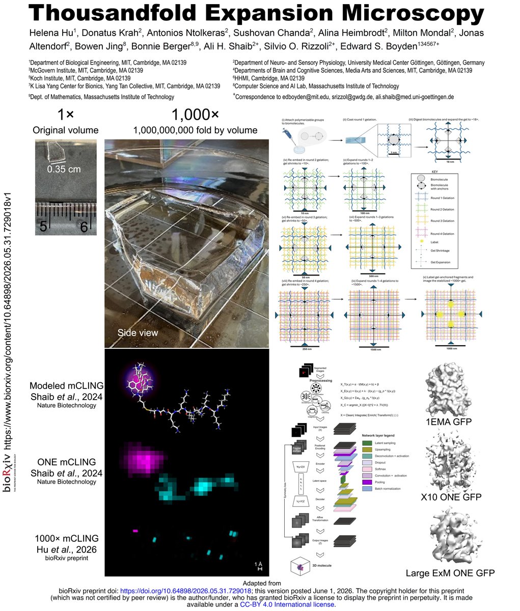
Hello world, meet 1,000× Expansion Microscopy.
1,000,000,000× expansion by volume! A gel that starts at a few centimeters will then expand to the volume of an Olympic swimming pool. https://t.co/E43kxx4O5M
In our new bioRxiv preprint, work carried out between MIT and UMG, led by Helena Hu in collaboration with scientists from the labs of @eboyden3 Ed Boyden, Silvio Rizzoli, and myself, we present Thousandfold Expansion Microscopy.
By enlarging biological specimens across multiple rounds of expansion, molecular-scale features, as small as the distances between adjacent amino acids, can be visualized with conventional optical microscopes.
Democratizing super-resolution microscopy.
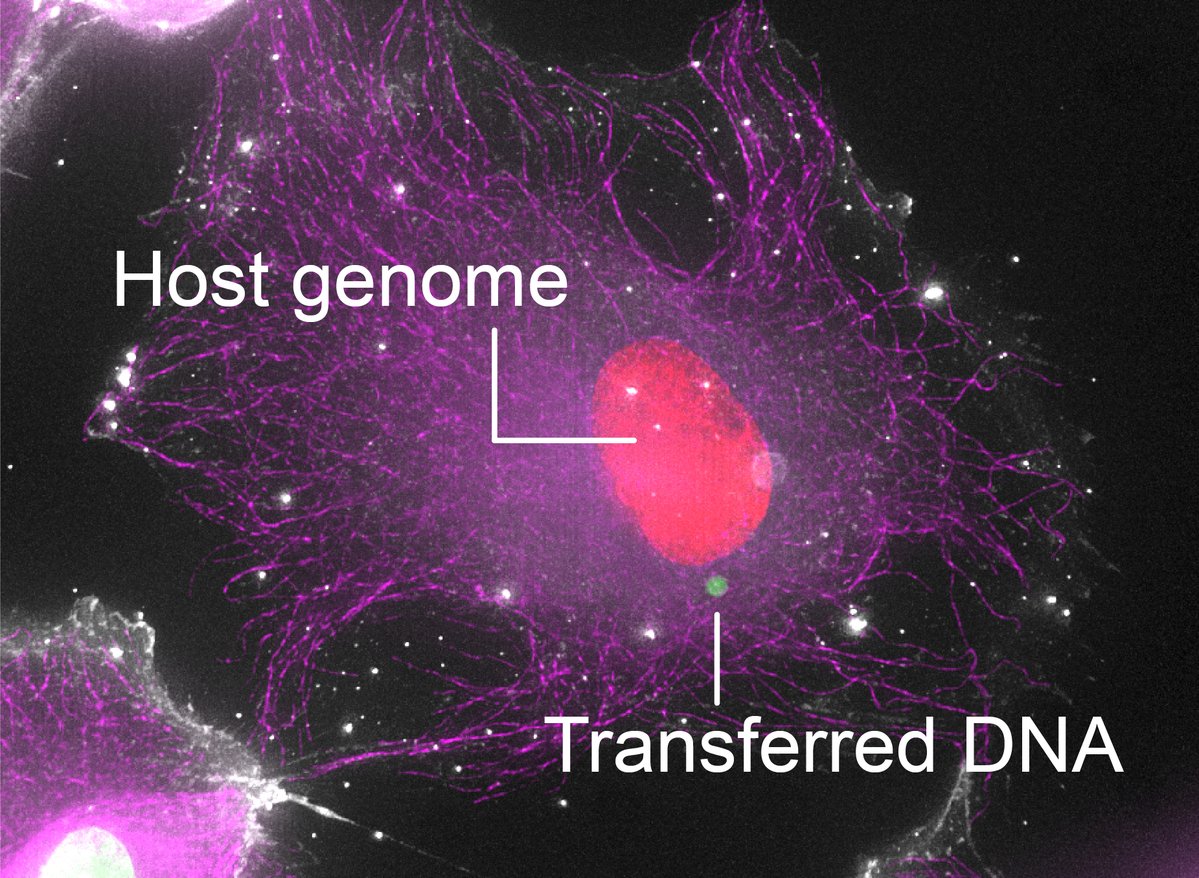
Excited to share our latest paper, out today @CellCellPress. We found that large pieces of the human genome can transfer between cells upon direct contact, endowing recipient cells with heritable phenotypic changes. (1/7)
https://t.co/SbshGhofN0
This work was a wonderful collaboration with Dr. Takada’s group. We’re grateful to all collaborators, especially our graduate student and co-first author Sakuntha Gunarathna (@galaxyhive), for his important contributions.
Excited to share that our new paper is out!
We analyzed cfDNA from patients with metastatic breast cancer and captured fragmentomics changes after CDK4/6 inhibitor treatment—offering a potential path to monitor/predict treatment response.
https://t.co/gQL8joTBlr
Excited to share our new paper in @NatureGenet led by the incredible @6jinLee! We uncover how cohesin loss impairs postmitotic refolding of 3D chromatin topology with limited context-specific effects on gene activation—especially during differentiation. https://t.co/sCPSDjAa8a
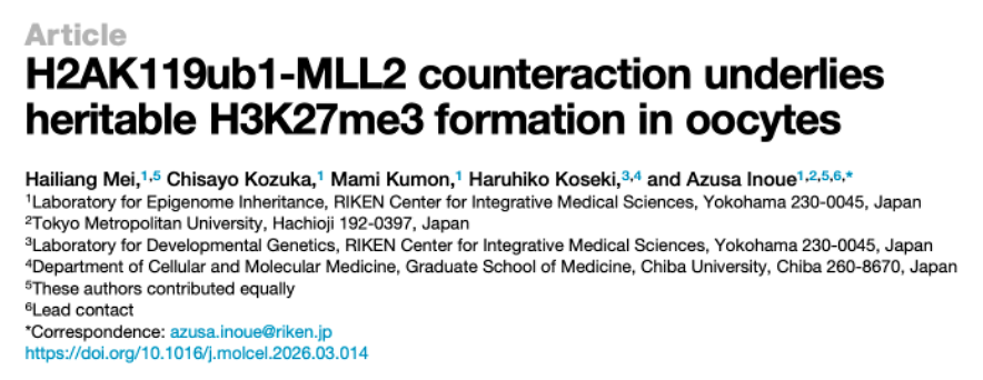
Hello Polycomb and genomic imprinting lovers!
Our new paper is out in @MolecularCell — we uncovered how H3K27me3 (also non-canonical imprinting) is established in mouse oocytes. This was made possible by generating oocyte-specific triple cKO mice!
Summary in thread↓↓ (1/10)
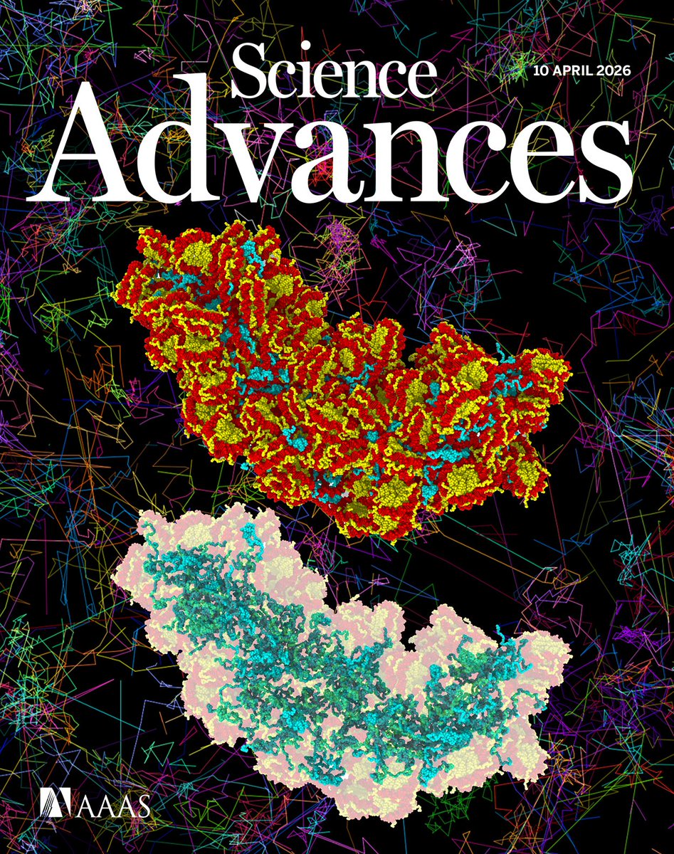
Our linker histone H1 paper is featured on the cover of the April 10 issue of @ScienceAdvances.
https://t.co/fGZsmvRPWK
A molecular dynamics simulation of nucleosomes and linker histone H1, with single-H1 trajectories (background). 🎉 Full cover caption: https://t.co/Cj4Ey5RRfs
I am excited to have been awarded a Wellcome Trust CDA fellowship. This award allows us to dissect the mechanistic basis for trophectoderm lineage commitment across scales, from atoms to embryos.
https://t.co/c1trXmdHH7
We are looking to hire a Postdoctoral Fellow to join the Eda Yıldırım Lab at Koç University through the Koç University Postdoctoral Researcher Support Program.
Please share with potential candidates in your network!
We develop cf-EpiTracing, a new liquid biopsy tl to trace tissue origin and therapy responses using cell-free multimodal chromatin states from as little as 50 ul plasma, now
@Nature. Congrats to Xubin, Xiaoxuan, etc. Big thks to many many collaborators! https://t.co/kLkbr1WlLb
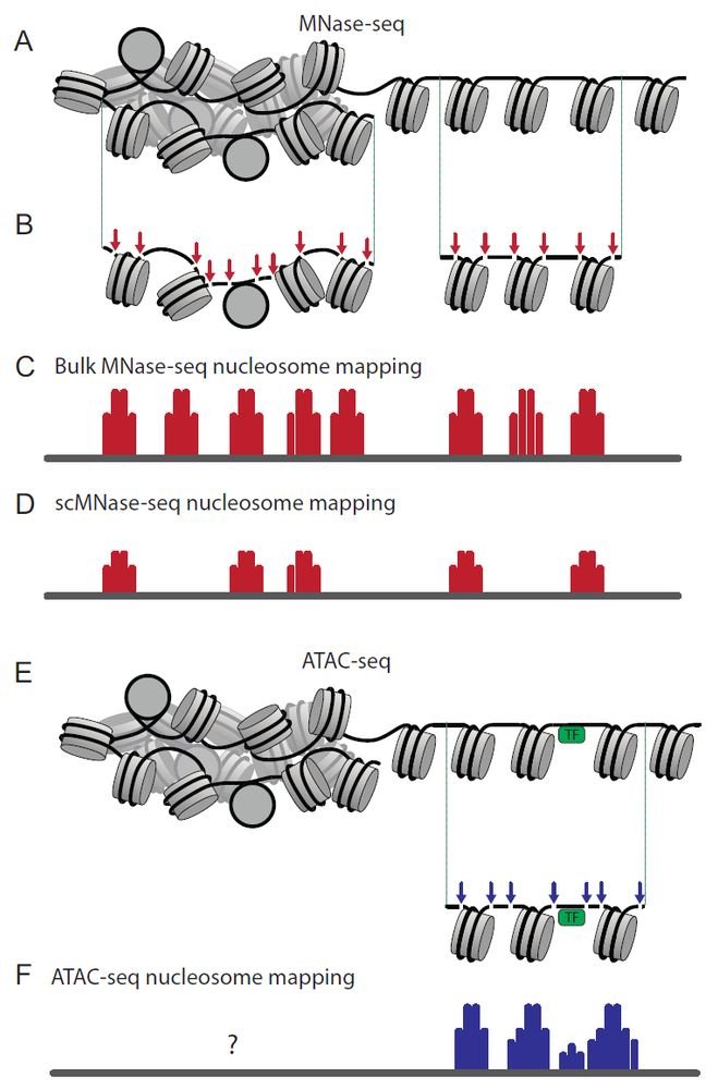
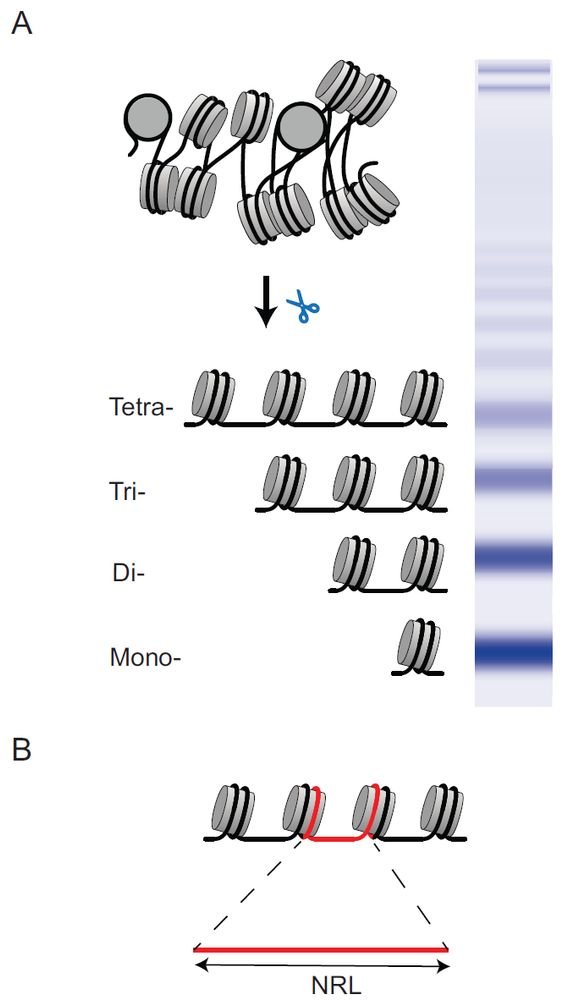
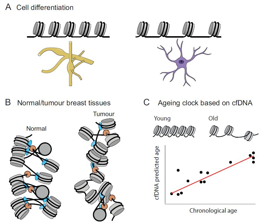
Nucleosome aficionados! Our new review "Nucleosome spacing across cell types, diseases, and ages" is out in NAR: https://t.co/Z1TgEYxBH8
A huge effort to pull together what we’ve learned about nucleosome spacing in many systems. Enjoy!
#nucleosomics#nucleosome#chromatin#cfDNA
🚨 Today in @Nature, we report GEMINI—a genetically encoded intracellular memory device that writes cellular dynamics into tree-ring-like fluorescent patterns within cytoplasmic protein assemblies.[1/n]
https://t.co/eVchPCiK6f
Our collaborative study with Yasui lab @sciencetokyo_ja is out in @ScienceAdvances ! With gRNA/GeNL tagging, we showed that cancer-derived small EVs can reach and be concentrated in urine via transcytosis, providing rationale for urinary liquid biopsy. https://t.co/4F23HMTuL7
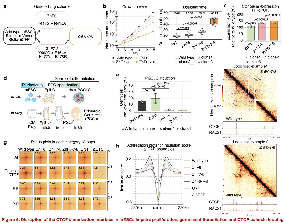
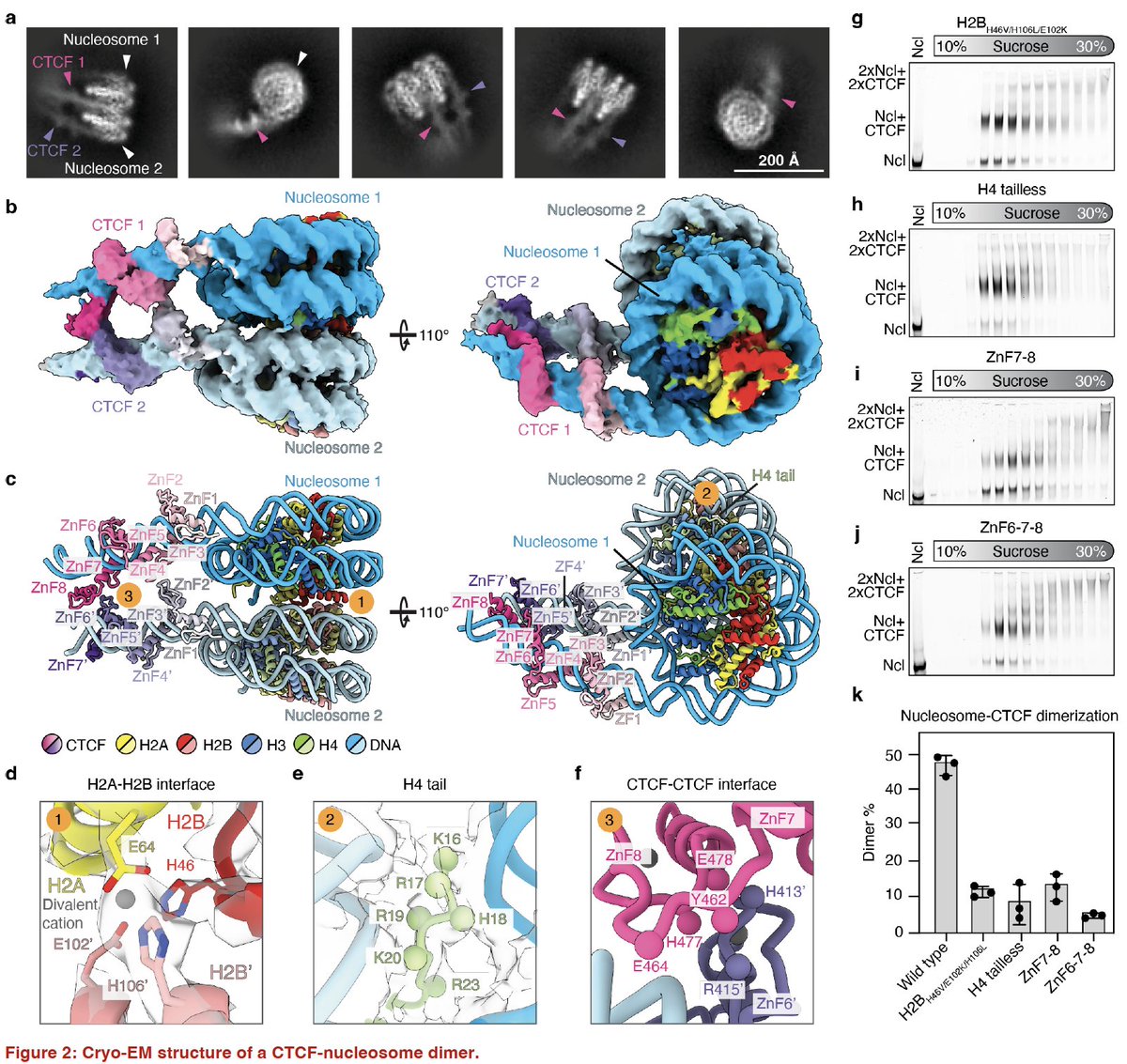
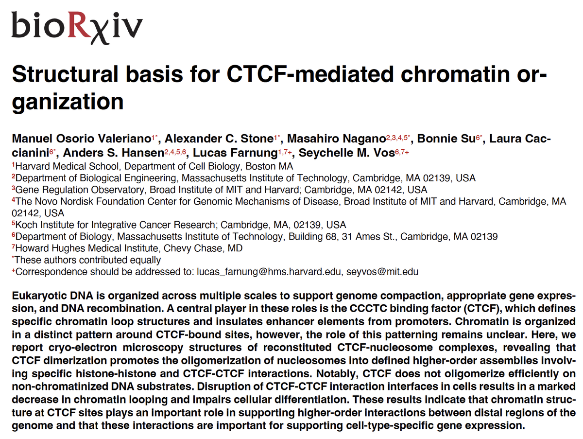
Masahiro Nagano and I were fortunate to collaborate with the Vos&Farnung labs:
CTCF dimerization drives higher order nucleosome assemblies.
Disrupting CTCF dimerization in cells weakens CTCF loops and causes growth + differentiation defects.
https://t.co/QLNtV0kvWD
My student @Aoi_Otsuka_’s PhD work is now officially published @CSF_JSCB!
🔬Single-nucleosome imaging uncovers biphasic local chromatin dynamics in an oncogene-inducible human carcinogenesis model (1–3 d: same, 5–7 d: ↑, back by week 4). Congrats! 👏
https://t.co/2XjYoMj7gv
Happy to see my last piece of postdoc work online—nearly two years after it first appeared on bioRxiv.
Title: Live-cell single-molecule dynamics of eukaryotic RNA polymerase machineries
https://t.co/StOMJzxzae


![DingchangLin's tweet photo. 🚨 Today in @Nature, we report GEMINI—a genetically encoded intracellular memory device that writes cellular dynamics into tree-ring-like fluorescent patterns within cytoplasmic protein assemblies.[1/n]
https://t.co/eVchPCiK6f https://t.co/KPmYKFgnZt](https://pbs.twimg.com/media/HCgxFtcWkAAoaPS.jpg)


