(1/8) My latest study is out in @Nature ! Kudos to coauthors especially @jess_cardin and to @KavliAtYale and @WuTsaiYale for support. We find that gamma power in mouse visual cortex is caused by brief events supporting visual processing https://t.co/h45HEg0LpX
The University of Arizona is holding an open-rank neuroscience faculty search, multiple tenure-track positions available! Positions will be in the new Department of Translational Neurosciences at the College of Medicine in Phoenix. https://t.co/G758sOFZ4S
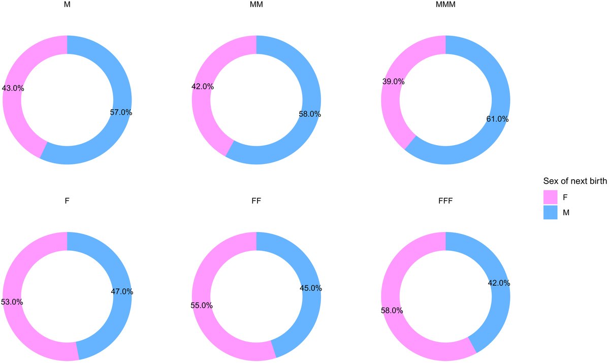
Sex at birth may not be purely random, according to a new @ScienceAdvances study.
Instead, each family’s likelihood of having only male or only female children is a “weighted coin toss” influenced by maternal age and genetics. https://t.co/msMvLfLuuz
Neuronal activity influences the growth of #myelin sheaths along axons by signalling through metabotropic glutamate receptor 5 (mGluR5) on oligodendrocytes
https://t.co/1GDKKMAgtZ
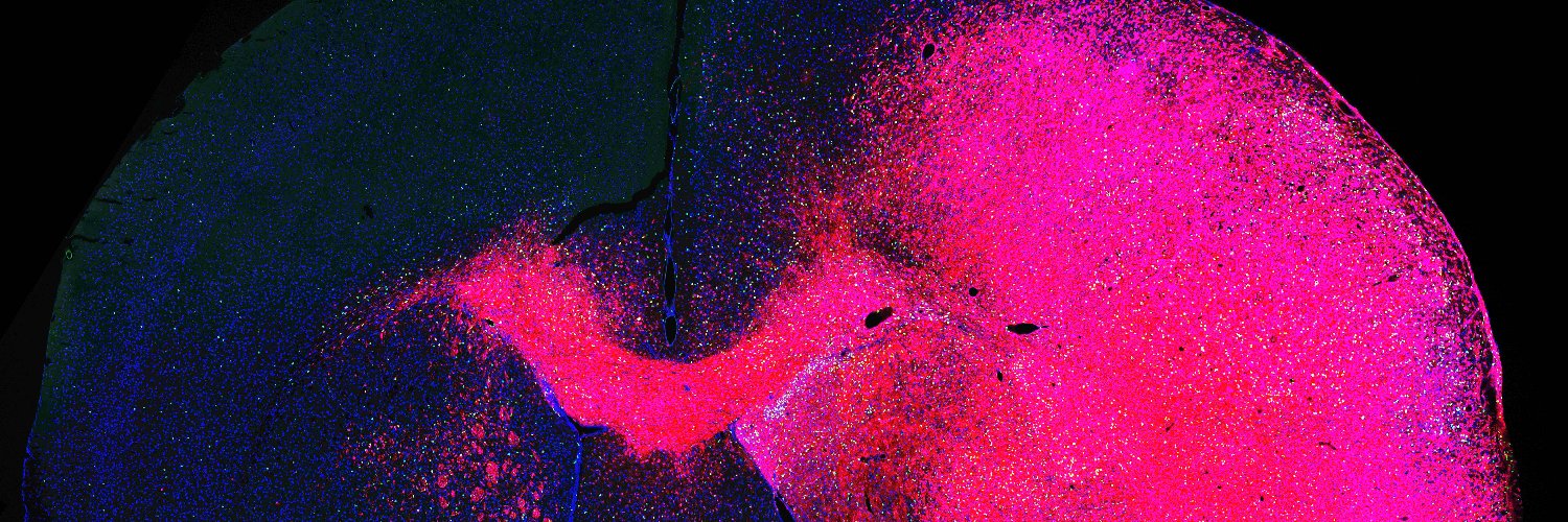
Today in @Nature, we report a spatial single-cell atlas of human cortical development, revealing surprisingly early specification of human cortical layers and areas.
We built an interactive browser to explore the spatial data: https://t.co/Xy3JcsSpYw
Paper link below👇
Thrilled to share our paper, New in @ScienceMagazine! We show that transiently boosting ketamine-induced ERK activity prolongs its antidepressant effects via enhanced synaptic plasticity and synaptogenesis in CA1. Grateful for all the support and guidance from mentors and colleagues @VanderbiltBrain & @VandyPharm along the way! @LisaMonteggia, Ege Kavalali, @natalieguzi, Ji-Woon Kim, etc.
Also, thank Dr. Hashimoto for the nice perspective on our paper.
https://t.co/ahign22dfM
https://t.co/dleZnN5zyJ
#scienceresearch
#ketamine #erk
#antidepressant
A new Science study finds that different dendritic segments of a single neuron follow distinct rules. The results challenge the idea that neurons follow a single learning strategy and offer a new perspective on how the brain learns and adapts behavior
📄: https://t.co/4eu4Rvw6NZ
#SciencePerspective: https://t.co/93KqQH1Ti5
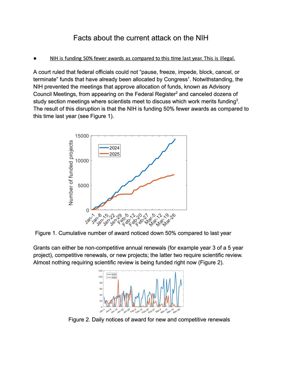
Scientists must push back against the threat of rising white nationalism and the dangerous and pseudoscientific ideas of eugenics
https://t.co/mpm0ijjCFm
The deadline to register for the Annual Southwest Developmental Biology meeting is fast approaching! Don’t miss out—register soon, and we’ll see you in New Mexico! More information about the program and speakers here https://t.co/AnPsOqZyIa
Glioblastomas hijack the normal brain circuitry for rapid proliferation. Fascinating @Nature study showed that malignant brain tumors, or gliomas, infiltrate and integrate across brain networks. Pharmacologic blockade of neuron-glioma interaction reduce tumor proliferation.
Whole-brain projections of over 7,000 hypothalamic neurons expressing distinct neuropeptides in male mice, identifying 2 main classes and 31 types using single-neuron projectome analysis
https://t.co/a3qTy1UfJ7
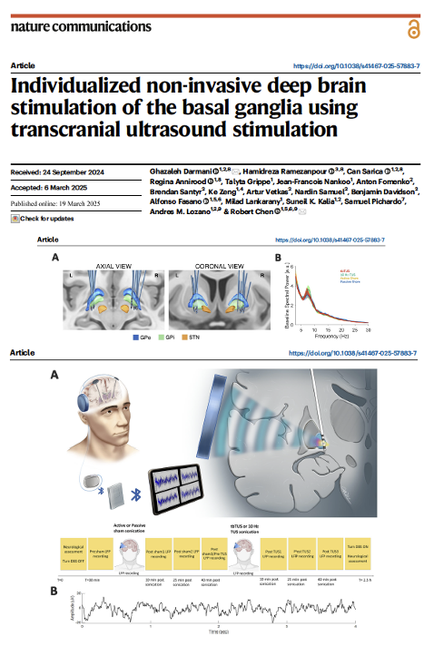
Wouldn't it be cool to be able to stimulate deep within the brain without the need for brain surgery (i.e. DBS), and without actually making a physical lesion in the brain (i.e. focused ultrasound or pallidotomy)? This paper provides 'proof of concept' that a transcranial ultrasound stimulation (TUS) approach may be achievable. Darmani, Chen and colleagues bring us up to speed in Nature Communications.
Key Points:
- The authors provide an interesting study that examined TUS-induced changes in 10 individuals w/ Parkinson’s and dystonia.
- There were also 15 healthy controls.
- Physiology and local field potentials were sampled from deep brain stimulation leads w/in the globus pallidus.
- Theta burst TUS increased theta power during DBS.
- The authors showed 10 Hz TUS enhanced beta power and the effect persisted for ~40 min.
- Interestingly, the GPi TUS actually prolonged stop-signal reaction times. There was also impaired response inhibition.
My take: What was interesting was that transcranial ultrasound was able to engage a common target (GPi) used for DBS surgery and focused ultrasound. We all remain 'deeply' interested in noninvasive deep brain stimulation as this could open the door for a safer and potentially (one day) a more practical approach for stimulating from 'outside the brain.' The field has a ways to go, but this is a nice early step. It is critically important that the authors were able to show us that TUS was able to 'modulate neural circuits in a spatially precise manner.' We must – respice finem – consider the outcome, when developing the less invasive methods of brain stimulation. I don't think we should be too disappointed with the early results, as this road will be a long but worthwhile journey. https://t.co/WE0F7A2mJT #parkinson #deepbrainstimulation #focusedultrasound @ParkinsonDotOrg@FixelInstitute@Nature@NatureComms@DBSThinkTank
HUGE congratulations to @IamLinghua & team for her latest @Cancer_Cell study
Integrative analyses of over 14 million cells from 10 cancer types across 7 spatial transcriptomics and proteomics platforms identifies four distinct spatial CAF subtypes conserved across cancer types