Exciting advancements in EP 3D anatomic imaging with ICE integration! Proud to share that @UCSDHealth recently achieved a First in Human milestone using the ViewFlex X SE catheter in a clinical study with @AbbottCardio for high-quality imaging. #EPeeps@TY_TACKacardia
Enjoyed assisting with some workflow and technology improvements in FIH trials of novel Viewflex X ICE catheter from @AbbottCardio -- we performed ablation of PVC from suspected intramural site with RF to septal RVOT and adjacent ASOV. Will be curious to see what @dhakalbish thinks with his @PennEPFellows based ICE approach in trials tomorrow. Contouring of key structures including pap muscles and ASOV shown below:
@AMatthews0@mikelean@Zachkoch13
Monday started with First-in-Human Use of “ViewFlex X SE” ICE Catheter with @AbbottCardio
I've always loved using the ViewFlex ICE for its exceptional image quality, but now with seamless ICE integration into Ensite X, this is a total game-changer that enhances precision like never before.
Some features that stood out to me
🔹 ICE Integration with Grid and FAM Mapping - Real-time visualization that aligns perfectly with 3D GridX and FAM maps. This precision ensures more efficient targeting of critical anatomy, improving procedural accuracy.
🔹 Smooth Transition to the Left Atrium
Despite its softer shaft, the ViewFlex X SE provided excellent maneuverability and control, allowing smooth navigation to the left atrium while maintaining consistent imaging quality.
🔹 Maintaining Outstanding Image Quality
For those of us who’ve always trusted the ViewFlex ICE platform for its superior imaging, this new catheter doesn’t disappoint—it adds seamless integration without compromising clarity.
#3DMapping #PrecisionMedicine #ViewFlexXSE #Innovation #EPWorkflow @StBernards #epeeps #ArrhythmiaResearchGroup
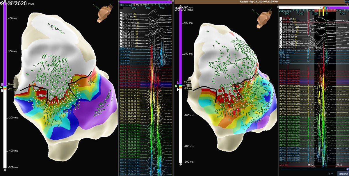
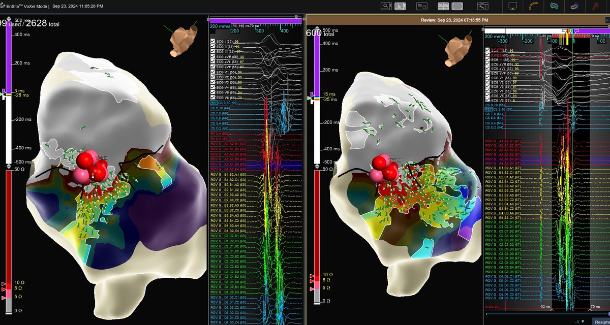
First burn termination of AVRT in <1 second with help from #HDGrid and OT Near Field.
AVRT and RV paced maps show clear breakout on the Mitral Valve. OT vectors & Peak Frequency overlay at 350Hz confirm site of termination.
Mapped by @ValayParikhMD@AbbottCardio
Correlation between VT isthmus, imaging findings and histopathology in a patient with desmoplakin cardiomyopathy.
A collaborative report with @ColumbiaPS pathology team published in @JACCJournals#JACCEP@WeillCornell
https://t.co/WsUSwL8j8r
Truly remarkable remote case support at HRS by Dr. Bernard, @Skhatib55, and our Abbott Remote Specialist team.
Really powerful demonstration of how #EnSiteConnect & @medinbox can transform the reach of EP.
#AbbottProud#HRS2023
A world’s first for treating the most common abnormal heart rhythm. Our TactiFlex Ablation Catheter, Sensor Enabled, is now FDA approved, making it a first-of-its-kind treatment option for atrial fibrillation. https://t.co/AfS2apVTsA
ISI: https://t.co/T3LK4KnfW1
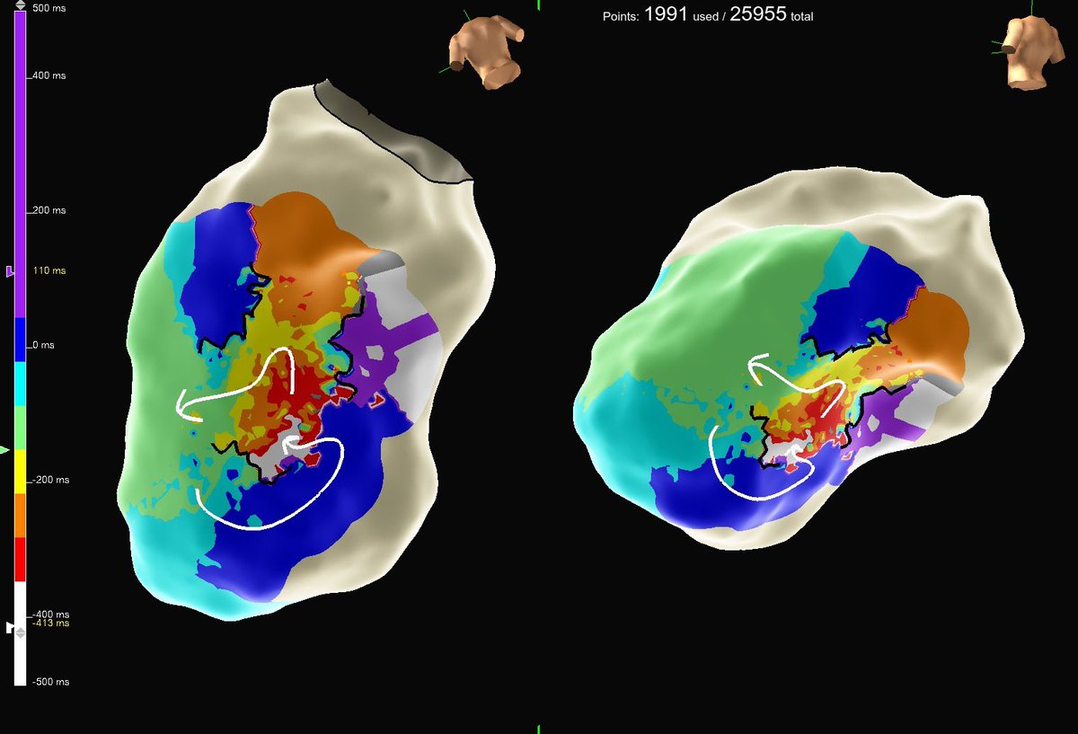
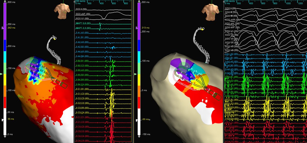
ECMO patient referred to EP lab for VT storm. RV paced ILAM guided ablation strategy with #HDGrid helped target areas of late activation and isochronal crowding. Patient is off ECMO and discharged from hospital.
Mapped by @JamesGabriels
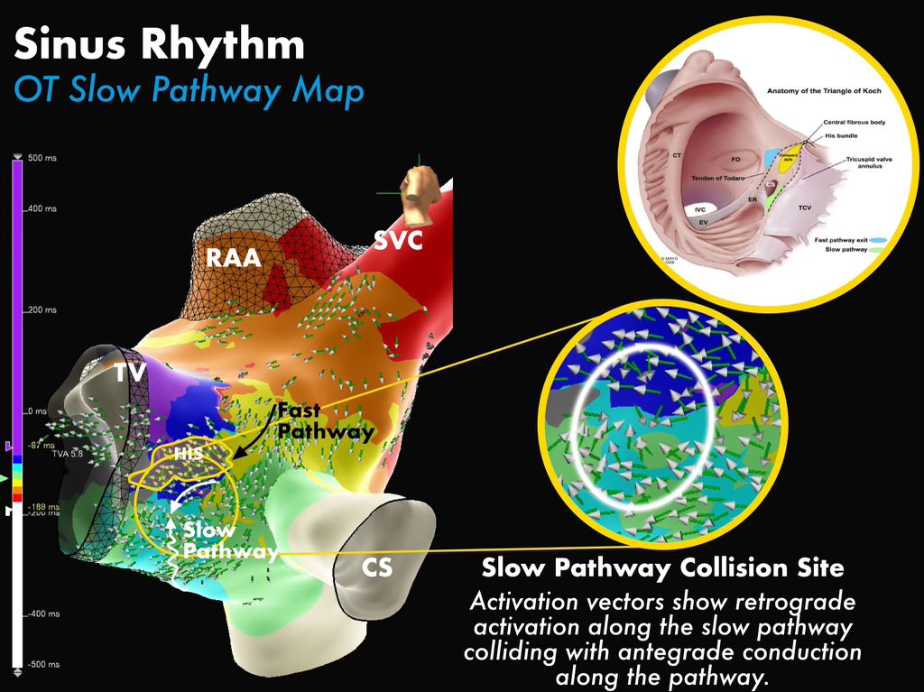
#EPeeps had a very fine day with #EnSiteX#HDGrid and #OT mapping slow pathway prior to #AVNRT ablation this week.
Observed nice slow junctionals with RFA at site of collision. Non-inducible post-ablation.
Maps by @MeganMalladiEP
Figures by @sota_ep
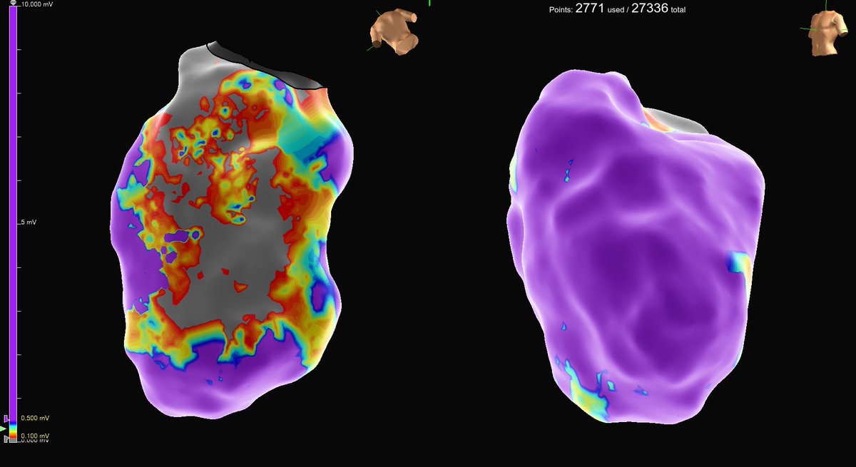
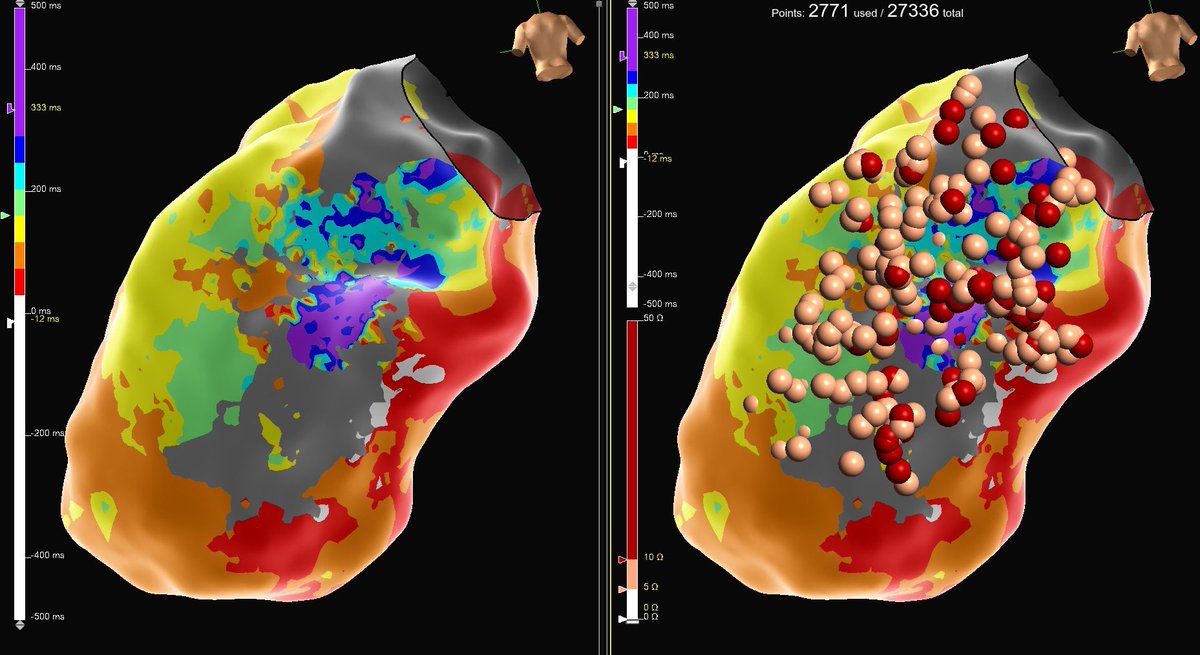
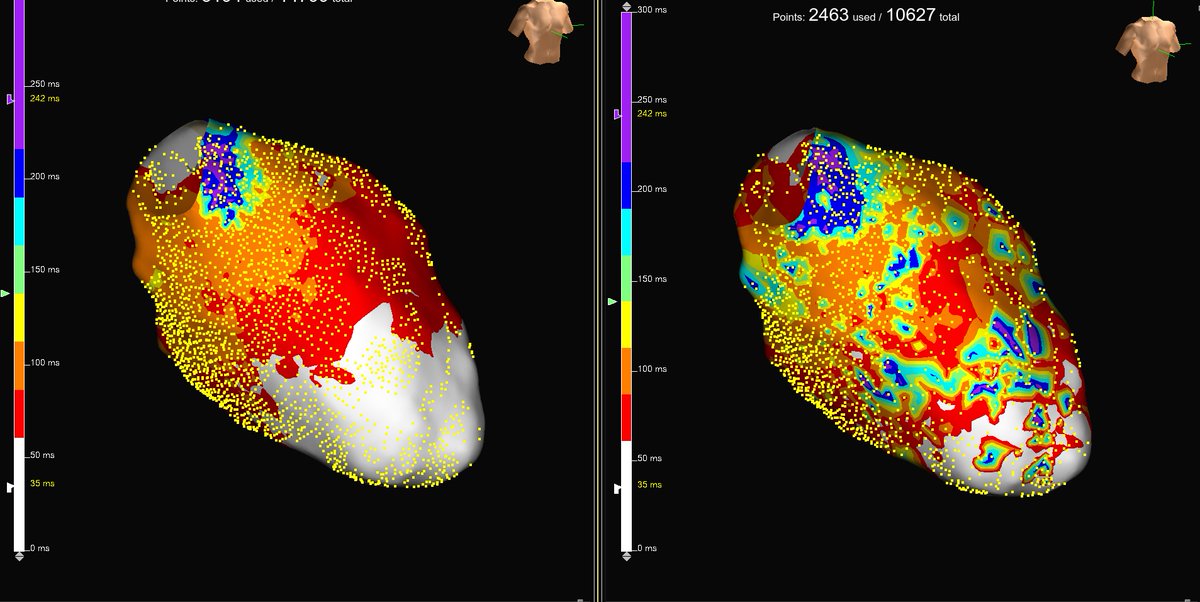
Omnipolar is game-changing for automated ILAM approach. New(L) vs. old(R) auto annotation display without any manual annotation. Yet another localized periaortic VT substrate from LMNA, with diastolic 🍽️ buffet during VT within sinus rhythm deceleration zone. @AbbottCardio