In a prion disease mouse model that phagocytic Gpnmb⁺ microglial state reflects regional responses to cell death and lysosomal stress rather than direct sensing of amyloid fibrils
@NatureComms@AdrianoAguzzi@Elena_DeCecco 🇨🇭
https://t.co/VsBnBOPc4g
@LukensJohnR Very interesting paper which very much aligns with our recent work on Gpnmb in neurodegenerative disorders (in our case prion disease) https://t.co/PTMN3jl9Uu
🚨New preprint from our lab!🚨
You've heard of CAR-T cell therapy for cancer. But have you heard of CAR-NSC for stroke?
8+ years of work, this may be the most important work of our research so far!
Read more 👇
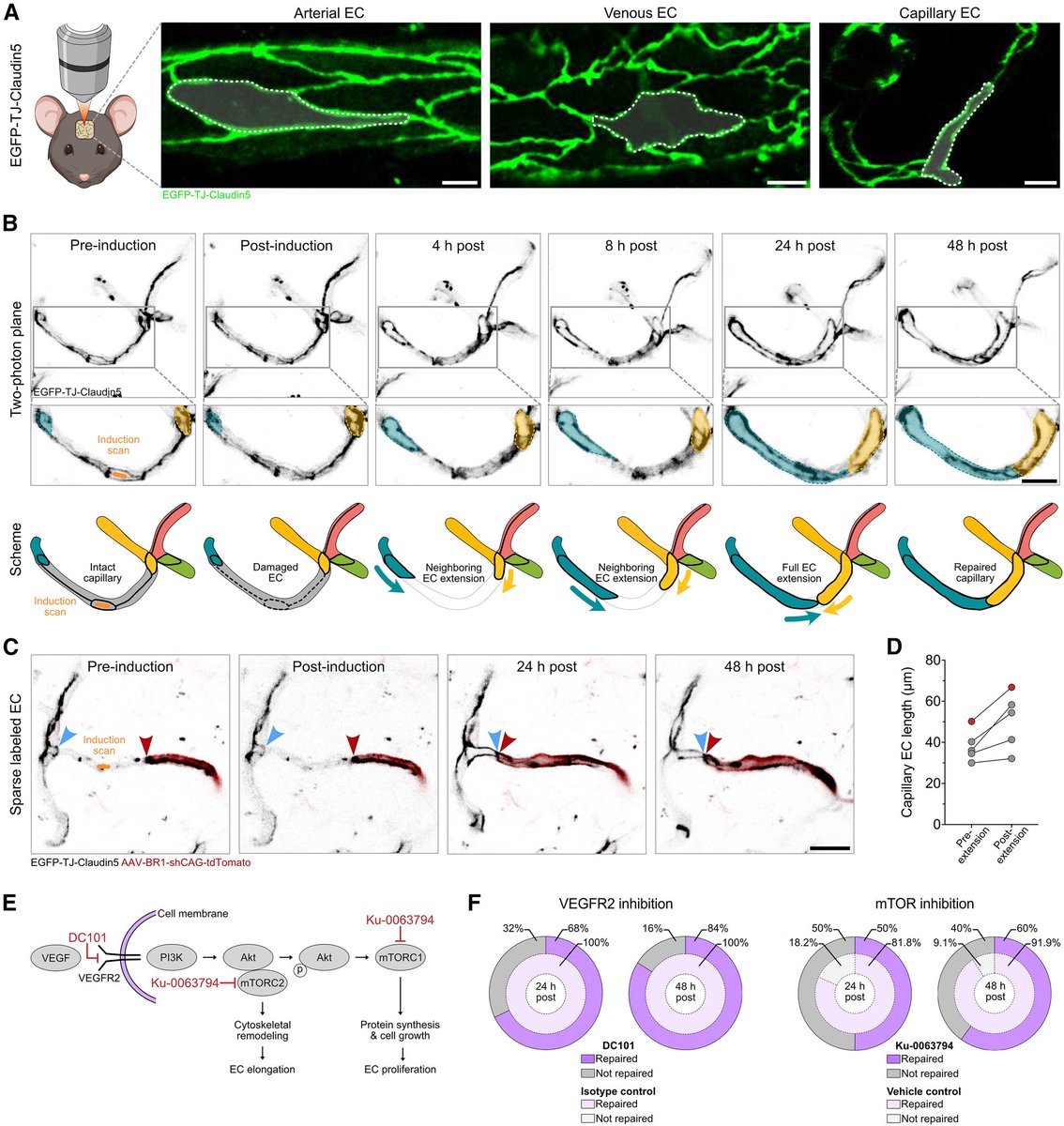
Did you know that if you remove a single endothelial cell from a brain capillary, neighboring endothelial cells have the capacity to rapidly extend to rebuild the vessel? Excited to share our new paper in Neuron. Congratulations to all co-authors. https://t.co/QsAtMH1598
Importantly, when we removed Gpnmb from microglia, their ability to phagocytose apoptotic cells was significantly impaired, pointing to a critical role for Gpnmb in microglial clearance mechanisms during neurodegeneration.
This microglial state was not present at earlier (but still late) timepoints. Stroke induced animal model proved that indeed cell death alone can trigger Gpnmb+ microglial state!
Excited to share our new paper in @NatureComms ! 🧠🔥
https://t.co/PTMN3jl9Uu
Using spatiotemporal transcriptomics and functional validation, we identify a Gpnmb⁺ phagocytic microglial state associated with cell death during prion disease progression.
#Prions#STtranscriptomics
Interestingly, we also observed Gpnmb⁺ microglia in white matter regions associated with demyelination, supporting the idea that this state may represent a broader phagocytic response to cellular damage.
Gpnmb⁺ microglia were not broadly distributed across the brain, but instead localized specifically to regions undergoing severe cell death, suggesting that the surrounding tissue environment — rather than prion aggregates alone — plays a major role in shaping microglial states.
Happy to see this work now published in PLOS Pathogens.
We identify TFAP2C as a functional interactor of hnRNPK and show how this axis regulates prion propagation via mTORC1 signaling.
Great team effort—congrats to Stefano Sellitto and all co-authors!
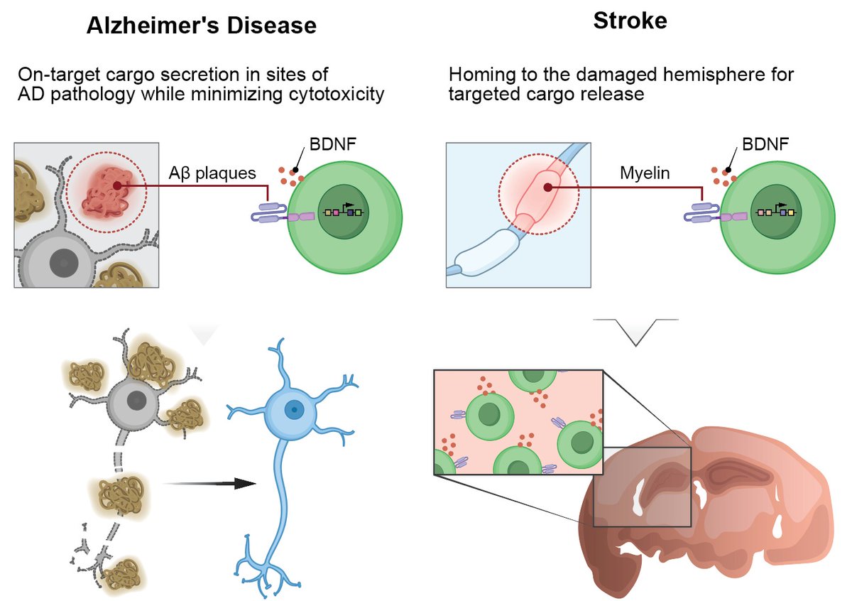
Excited to share two back-to-back studies with @jonykipnis@boskovic_p showing the promise of CAR T beyond cancer — in Alzheimer’s and stroke — inspired by the visionary work of Zelig Eshhar.
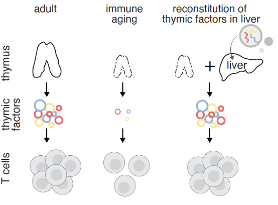
Excited to share our new work on immune aging! We explored whether the liver could serve as a temporary "factory" to produce immune factors that decline with aging, potentially helping to rejuvenate aged immunity. @mircoscopy.
We have now reached a new level of spatial omics: translatomics! https://t.co/GXEtIhrpKd Fascinating new tech in new paper "Scalable spatial single-cell transcriptomics and translatomics in 3D thick tissue blocks" from @naturemethods
Very elegant work from an amazing Italian scientist @SimonaParrinello!
Axonal injury → Wallerian degeneration → neuroinflammation → glioma growth. And it’s targetable.
#Glioma#Neuroinflammation#BrainCancer#Nature
Congratulations to all the author of this beautiful work!
🧬 BREAKING: Our CRISPR-GPT paper is out TODAY in Nature Biomedical Engineering @natBME !
🤯 We built an AI agent that turns ANYONE into a gene-editing expert in 1 DAY instead of months. An undergrad with ZERO experience achieved 90%+ editing efficiency on their FIRST attempt.
🧵 Here's how we're building expert AI agents for cutting-edge biotechnology:
🎯 The Problem: CRISPR is revolutionary but requires PhD-level expertise, it can take weeks to learn, adopt, and design, analyze a CRISPR experiment for R&D or making life-saving medicine. Even Pro scientists can make small mistakes (e.g. typos in guideRNA or cloning design) that cost months to find out, slowing us down.
💡 Our Solution: CRISPR-GPT - an AI co-pilot from @Stanford@Princeton@GoogleDeepMind that guides you through EVERY step via simple conversation
🔬 Real Results:
-Novice researcher: ~90% editing on 1st go
-Training time: Months → 1 day
-100% success rate in our trials
-Even experts save days/weeks on data analysis & troubleshooting
🤖 How it works: Our multi-agent system handles: CRISPR system and delivery method selection, guideRNA design, Protocol generation, Real-time troubleshooting, Data analysis, and beyond. All through natural language! No coding, no complex software.
📊 We benchmarked it extensively:
-288 evaluation scenarios/cases
-Outperformed GPT-4o on ALL gene editing tasks
-Trained on 11 years of expert discussions
-Covers knockout, base-editing, prime-editing & epigenetic editing
🌍 Why this matters:
-Every lab can now use CRISPR with an AI system distilling expert knowledge and skills.
-Every student can learn faster.
-Every researcher can tackle bigger challenges without worrying about small mistakes.
-Customized CRISPR design can be automated based on your need and the context of R&D workflow.
-Agentic AI ensure safety, privacy, and responsibility
-We're not just automating gene editing - we're using AI to power scientists to cure diseases.
🚀 Try it yourself! Beta access available at: https://t.co/CFQzrdVVPW
Paper: https://t.co/WOgmAjg1Ed
Code: https://t.co/UnPKrxyJuW
Benchmark (companion work, Genome-bench): https://t.co/LIooAXj2op
Co-first and key authors: @YuanhaoQ@KaixuanHuang1@MingYin_0312
PIs: @lecong@MengdiWang10
Key collaborators: @Rbaltman@denny_zhou
The future of biology and science is conversational. The future is now. @natBME@NaturePortfolio
#CRISPR #AI #GeneEditing #Biotech #Science #AISafety
In this study, we generated a Slco1c1-CreERmouse line (Slco1c1-KI-P2A-iCreERT2, briefly, Slco1c1-CreERKI). Our staining results validate its high specificity for labeling CNS endothelial cells, with rare expression in peripheral endothelial cells.
https://t.co/dfIk6wvLRT