The danger of reasoning by analogy: "The behavior of things on a very tiny scale is simply different. (Atoms) do not behave just like particles. The do not behave just like waves. Atoms do not behave like weights hanging on a spring and oscillating. Nor do they behave like miniature representations of the solar system with little planets going around in orbit. Nor does it appear to be like a cloud or fog of some sort surrounding the nucleus. It behaves like nothing you've seen before."
Likewise, the cell is not a deterministic machine or factory. I think I can safely say that nobody understands the cell.
Any1 is a #phospholipid scramblase involved in #endosome biogenesis. From Jieqiong Gao, Christian Ungermann and colleagues: https://t.co/nBhHMI0Ihi
📕 In #Lysosomes 2026: https://t.co/4ANhoABM3P
Advanced Instrumentation Training Program (AITP)
Learn • Explore • Innovate
Hands-on Training with High-End Instrumentation
Open for Students, Researchers & Professionals
Certification with Practical Research Exposure
@iitdelhi@Pratishtha_DST@IndiaDST@Icosahedral16
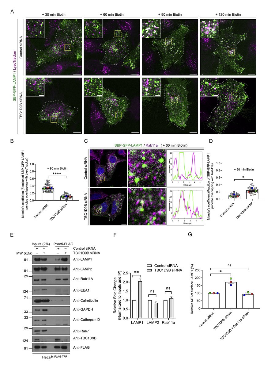
.@PriyaChauhan97, Yogita Phogat, @MahakSharma23 et al. report that the small GTP-binding protein Arl8b recruits the Rab11a GAP, TBC1D9B, to inactivate Rab11a-mediated recycling of newly synthesized LAMP1 and mediate its efficient sorting to #lysosomes. https://t.co/5HtOZkV8zj
Chandramouli Mukherjee @MukherjeeCm_02, Bhavani Shankar Sahu @urssahu@DBT_NBRC and colleagues find that Chromogranin B plays an essential role in post-ER sorting of dense-core vesicle cargo.
https://t.co/VftEF8mOkH
Kim, @spencerSK1 et al describe a mechanism for lysosomes to sense and respond to acute increases in their #membrane tension by relieving hydrostatic pressure via the outward transport of major osmoticants https://t.co/YHBykUIVCb
📕 In #Lysosomes 2026: https://t.co/LKzATqPq0I
Happy to have contributed to this preprint with Fangzhu Zhao and @realJimWells! LRP8-targeted bispecific antibody degraders degrade the selenium uptake receptor LRP8, reduce GPX4 / selenoproteins, and sensitize cancer cells to ferroptosis.
https://t.co/Ue9HCnnyrI
Proud to hood Sydney Tomlinson as she earned her PhD in Metabolic Biology at UC Berkeley. Sydney’s dissertation uncovered mechanisms for induced degradation of ER membrane proteins and advances our understanding of ER quality control.
Great to see this Spotlight article in Trends in Cell Biology highlighting our recent identification of a lipid droplet quality control pathway in which FSP1 protects PUFA-rich neutral lipids from peroxidation, thereby suppressing ferroptosis.
https://t.co/oumG9roaOh
Golgi–Mitochondria Contact Sites: The Hidden Lipid–Stress Interface
Mitochondria are no longer “stand-alone powerhouses.” They operate as networked organelles, physically coupled to the ER, Golgi, lysosomes, lipid droplets, and plasma membrane through membrane contact sites.
One emerging axis is especially underexplored: Golgi–mitochondria communication.
Recent reviews and mechanistic studies suggest that Golgi-derived membranes and lipids may participate in mitochondrial remodeling, respiratory adaptation, and stress signaling—not simply through vesicular trafficking, but via direct or three-way ER–Golgi–mitochondria contact platforms. The Golgi contact-site review The Fast and the Furious: Golgi Contact Sites highlighted Golgi–mitochondria contacts as one of the most experimentally supported new Golgi contact interfaces, especially in lipid exchange and mitochondrial dynamics.
The key concept:
Golgi-derived PI4P/lipid-enriched vesicles may be recruited near ER–mitochondria contact sites, creating a three-organelle signaling hub that coordinates lipid supply, mitochondrial fission, and metabolic adaptation. This shifts the Golgi from “post-ER cargo station” to an active regulator of mitochondrial state.
A second layer comes from COPI biology. COPI is classically known for Golgi–ER retrograde trafficking, but a 2023 Cell Reports study showed that COPI disruption reduces mitochondria–ER contact sites, impairs Ca²⁺ handling, increases mitophagy, decreases respiratory capacity, and accelerates axonal degeneration. Restoring MERCS partially rescued mitochondrial and neuronal phenotypes.
This suggests a causal chain:
Golgi–ER traffic → MERCS integrity → mitochondrial Ca²⁺/respiration → cell survival
For aging, neurodegeneration, cancer metabolism, and fibrosis, this is a rich hypothesis space. Golgi–mitochondria/MERCS dysfunction could act as a hidden “organelle logistics failure” linking lipid imbalance, mitochondrial fragmentation, impaired respiration, and chronic stress signaling.
The field now needs better tools: split-FP proximity reporters, EM tomography, proximity labeling, lipidomics, and perturbation screens to distinguish true contact-site biology from nearby organelle crowding. A 2023 methods review provides a useful technical roadmap for studying membrane contact sites.
Working hypothesis:
Golgi–mitochondria contact is not a minor cell-biology curiosity. It may be a tunable metabolic control node—where lipid trafficking, mitochondrial dynamics, and disease stress programs converge.
Key references
David Y, Castro IG, Schuldiner M. The Fast and the Furious: Golgi Contact Sites. Contact. 2021. DOI: 10.1177/25152564211034424.
Maddison DC et al. COPI-regulated mitochondria-ER contact site formation maintains axonal integrity. Cell Reports. 2023. DOI: 10.1016/j.celrep.2023.112883.
Diokmetzidou A, Scorrano L. Mitochondria–membranous organelle contacts at a glance. J Cell Sci. 2025. DOI: 10.1242/jcs.263895.
Sarhadi TR, Panse JS, Nagotu S. Mind the gap: Methods to study membrane contact sites. Experimental Cell Research. 2023. DOI: 10.1016/j.yexcr.2023.113756
Explore fluorescent signals beyond the spectral options. TauContrast in STELLARIS Cryo allows you to exploit difference in fluorescence lifetime to separate overlapping fluorophores and identify unwanted signal contributions such as grid reflection. https://t.co/fh4Az4wMR2
We successfully concluded our One-Day Open House on Cell Biology & Advanced Microscopy Facilities at Central Research Facility (CRF), IIT Delhi – SATHI Foundation on 22nd April 2026.
@iitdelhi@IndiaDST@Pratishtha_DST