👉👉👉 A summary slide detailing MRD responses at approx 1 yr of combination (doublets and triplets) at different MRD thresholds in Firstline CLL @UTMDAnderson
We just reported our experience with steroid or anakinra prophylaxis in patients with high ferritin/CRP receiving axi-cel for DLBCL 1/n
https://t.co/9h2x340z1Q
🧬 #ScienceSaturday
What if some cancer-fighting T cells aren’t exhausted but simply waiting to be activated?
A new study published in @Cancer_Cell looked at T cells in the bone marrow of patients with multiple myeloma and acute myeloid leukemia (AML) to better understand how they recognize and respond to cancer.
➡️ Researchers identified a small group of tumor-reactive T cells that could recognize cancer cells. But unlike the exhausted T cells commonly found in solid tumors, these cells still showed strong cancer-killing features.
In other words, they appeared to be armed and capable of fighting cancer, but not fully activated.
➡️ The researchers also developed a 15-gene signature called TFiT that can help identify these tumor-reactive T cells.
When they looked at patients receiving immune-based treatments, including a bispecific antibody in multiple myeloma and immunotherapy in #AML, patients who responded were more likely to have these T cells present or see them expand during treatment.
Interestingly, the signature did not predict response to standard chemotherapy in AML, suggesting it may specifically reflect the immune system’s role in controlling the #cancer.
➡️ The team also found that many of the targets recognized by these #TCells did not come from traditional cancer mutations. Instead, they came from unusual protein fragments produced by cancer cells, and some were shared among multiple patients.
🧠 Why this matters: These findings suggest that T cells in bone marrow cancers may behave very differently from those inside solid tumors. Rather than needing to “wake up” exhausted T cells, some treatments may work by activating cancer-fighting T cells that are already there and ready to respond.
✨ Big takeaway: Understanding which T cells are already capable of recognizing cancer, and how to activate them, could help researchers develop more precise immunotherapies for multiple myeloma, AML and potentially other blood cancers. @mircoscopy@DKFZ@UniHeidelberg@broadinstitute@MIT
Read more here: https://t.co/RBzO2Mgxxn
1/ Our comprehensive evaluation of outcomes after anakinra treatment for ICANS, conducted in collaboration with our colleagues at OHSU, is out in @BloodPortfolio.
https://t.co/L89MQZ7nWT
🧱 This Jenga tower explains how venetoclax kills cancer cells.
In Jenga, each player pulls a block. The tower holds. Then one player pulls THE one block too many — and everything collapses.
Your cells work the exact same way. Every cell in your body carries a built-in self-destruct program: apoptosis. This program is ready to fire at any moment. What stops it? Protein-blocks holding the tower up. Here's the game:
🔸 The tower = mitochondrial potential
The mitochondrial membrane maintains a tension, an electrical potential. As long as the tower stands, the cell lives. If the tower collapses — if the potential collapses — cytochrome c escapes, caspases activate, and the cell dies. Irreversibly.
🔸 The stabilizing blocks = anti-apoptotic proteins (BCL-2, MCL-1, BCL-XL)
These proteins sit on the mitochondria like Jenga blocks. They restrain the killer proteins (BAX, BAK) and keep the tower from falling. More blocks, more stable tower. Less vulnerable cell.
🔸 The blocks being pulled = BH3-only proteins (BIM, BAD, NOXA, PUMA)
With every stress — DNA damage, survival signal deprivation, chemotherapy — the cell produces these small BH3-only proteins that neutralize a stabilizing block. One by one, blocks are removed. The tower starts to wobble.
🔸 Venetoclax = the player who pulls the BCL-2 block
Venetoclax is a specific BCL-2 inhibitor. It doesn't touch MCL-1. It doesn't touch BCL-XL. It targets one single block — and pulls it out.
If the tower was mainly held up by BCL-2 → it collapses → the cell dies. This is CLL, where BCL-2 is often THE load-bearing block.
If the tower was held up by MCL-1 → pulling BCL-2 changes nothing → the cell resists. This is one mechanism of venetoclax resistance in AML and some lymphomas.
And this is where it gets exciting.
BH3 profiling is a functional assay that measures, BEFORE treatment, how wobbly the tower already is — and above all, WHICH block is holding it up.
→ Very unstable tower ("primed" cell) = high BCL-2 dependence = venetoclax will work
→ Solid tower, stabilized by MCL-1 = a different strategy is needed
We're no longer just looking at mutations. We're testing the physics of the tower. Playing Jenga in vitro before playing it on the patient.
Next time you see a Jenga tower wobble, think of your mitochondria. They're playing the same game. All the time.
#venetoclax #apoptosis #BCL2 #BH3profiling #CLL #leukemia #hematology #oncology #MedTwitter
🧬 Why do some CAR T cells persist for years while others quickly disappear?
A new study uncovers a fundamental mechanism that may help explain long-term CAR T-cell durability: asymmetric cell division.
Although current FDA-approved CAR T products use either CD28 or 4-1BB costimulatory domains, these signaling modules produce markedly different clinical behaviors. Patients treated with 4-1BB-based CAR T therapies often experience greater long-term persistence, but the biological basis has remained unclear.
Key findings:
🔹 4-1BB CAR T cells generated daughter cells with distinct fates after their first division:
• One daughter became more effector-like
• The other adopted a more memory-like, persistence-prone state
🔹 CD28 CAR T cells, despite exhibiting greater surface protein asymmetry, showed less divergence in their transcriptional, epigenetic, and metabolic programs between daughter cells.
🔹 Multi-omic analyses revealed that costimulatory domains influence how early fate decisions are established, ultimately shaping whether CAR T cells become short-lived effectors or long-lasting memory cells.
Why does this matter?
Long-term remissions after CAR T therapy likely depend on a small subset of infused cells acquiring a durable memory-like phenotype. Understanding how costimulatory domains influence these earliest cell fate decisions could inform the design of next-generation CAR T products with improved persistence and more durable responses.
This study provides a mechanistic link between CAR design and long-term therapeutic success—highlighting that the future of cellular therapy may be determined by what happens during a CAR T cell’s very first division.
#CARTCellTherapy #CellTherapy #Immunotherapy #CancerResearch #Hematology #Oncology #TranslationalResearch #TCells #PrecisionMedicine #CAR_T
https://t.co/P6m8XlSmHA
CONGRESS | #EHA2026 | PRESENTATION
Nirav N. Shah presents phase I/II data on dual-targeted anti-CD20/anti-CD19 LV20.19 CAR T cells in patients with R/R CLL (N = 19).
Manufacturing was successful in 19/20 enrolled patients. The ORR was 90%, including CR/CRi in 79% and PR in 11%. With a median follow-up of 27 months, the 24-month PFS and OS rates were 77% and 83%, respectively, and all evaluable patients with MRD assessment (n = 8) became MRD negative between Days 30–90. CRS occurred in 95% (Grade 3–4, 11%), ICANS occurred in 26% (Grade 3–4, 11%), and IEC-HS occurred in 53% (Grade 3–4, 11%); mean ferritin levels were higher in patients with vs without IEC-HS (31,372 vs 1,162 ng/mL). After 2 DLTs at 2.5 × 10⁶ cells/kg, the dose was reduced to 1 × 10⁶ cells/kg. Expansion, ORR, and CR were similar by dose, while no Grade 3–4 ICANS or treatment-related mortality within 30 days occurred at the lower dose. CAR T-cell expansion was comparable between patients with vs without IEC-HS, but patients with IEC-HS had higher cytokine levels, including G-CSF, IFN-γ, IL-1ra, IL-2, IL-33, and IL-10.
Follow our live feed for more updates: https://t.co/SucPqSTSDg
Intended for HCPs only. This congress coverage is independently supported by pharmaceutical companies, who are allowed no influence on the content; a full list of supporters can be found on our website.
#lymphoma #lymsm #MedicalCongress @niravshahmd@MedicalCollege
Informative molecular analyses from TRIANGLE trial in mantle cell lymphoma, showing that the benefit for ibrutinib arms is driven by the TP53mut cohort.
Promising outcomes for TP53mut disease treated w TRIANGLE (2yr PFS ~75%)
?comparable to BOVen?
#EHA26#EHA2026#lymsm
CONGRESS | #EHA2026 | PRESENTATION
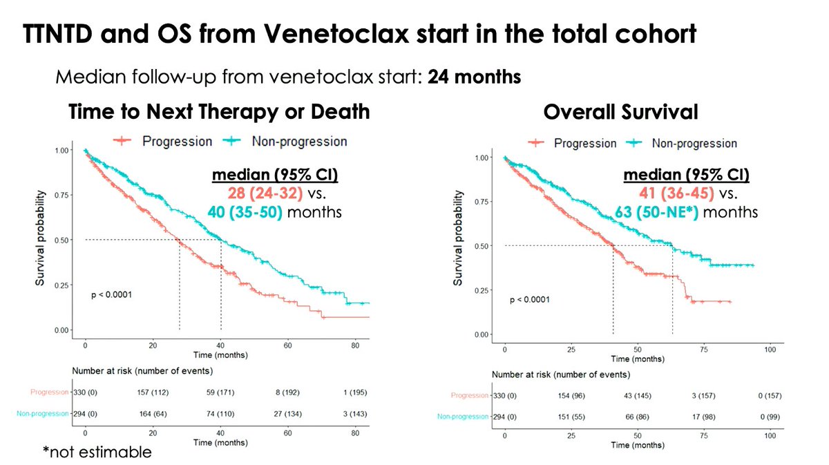
Thomas Chatzikonstantinou presents on an international ERIC study evaluating outcomes with venetoclax-based treatment after prior BTKi therapy in patients with CLL (N = 624; BTKi stopped for CLL progression, n = 330; BTKi stopped for non-progression, n = 294).
At venetoclax start, 45% received monotherapy, 37% received venetoclax + rituximab, 13% received venetoclax + obinutuzumab, and 6% received other combinations. After a median follow-up of 24 months, median TTNTD and OS in the total cohort were 33 months and 46 months, respectively. Patients stopping BTKi for progression had shorter TTNTD (28 vs 40 months; p < 0.0001) and OS (41 vs 63 months; p < 0.0001) vs those stopping for non-progression reasons. In multivariable analysis of the total cohort, non-progression as the reason for BTKi discontinuation was associated with longer TTNTD (HR, 0.68; p = 0.015) and OS (HR, 0.62; p = 0.009), while venetoclax + obinutuzumab was associated with longer TTNTD (HR, 0.46; p = 0.049) and OS (HR, 0.36; p = 0.047) vs venetoclax monotherapy. Male sex was associated with longer TTNTD (HR, 0.73; p = 0.037), while TP53 disruption was associated with shorter TTNTD (HR, 1.41; p = 0.041) and older age was associated with shorter OS (HR, 1.02; p = 0.016).
Follow our live feed for more updates: https://t.co/SucPqSTSDg
Intended for HCPs only. This congress coverage is independently supported by pharmaceutical companies, who are allowed no influence on the content; a full list of supporters can be found on our website.
#lymphoma #lymsm #MedicalCongress @INAB_Institute@CERTHellas@Ericllorg
Does HD-MTX ppx reduce CNS relapse even in ultra-high-risk LBCL? #lymsm
- >1900 pts, all CNS-IPI 5-6; testicular, renal/adrenal, breast; ≥3 EN sites
- 3-yr CNS relapse: 9.3% (no HD-MTX) v 8.1% (HD-MTX), adjusted HR 1.13 [0.82, 1.57]
- no diff in isolated CNS relapse: 5.9% v 5.7%
- no diff in relapse rates for any high-risk subtypes
- DHL/THL not included
End of HD-MTX ppx, although DHL/THL is still a question. cc @tobyeyre82@mattwilson2287
https://t.co/80zAeJmlXo
I recorded a video with @Medscape@MedscapeOnc during #ASCO26 on how to critically think about cancer clinical trials. It’s now out. Check it out and apply these principles when you’re evaluating any clinical trial presentation.
https://t.co/MdmWlBuoql
To help navigate the exciting content being presented at #EHA2026, the Lymphoma Hub Steering Committee members have provided their recommendations for the top abstracts to look out for in lymphoma and CLL.
Discover the list here: https://t.co/J8wKtcfDlO
#lymphoma#lymsm#leusm #MedicalCongress
van Rooij et al: MYD88 ddPCR on CSF cfDNA for non-invasive diagnosis of CNSL:
- 50 pts with confirmed PCNSL/SCNSL
- 203 with non-CNS lymphoma
- In combination with IL10/CXCL13, liquid biopsy had Sn 54% & Sp 100%
- Median corticosteroid to LP time 13 days
https://t.co/P9S5XMR8o5
During induction chemo for AML, why do we give continuous IV cytarabine, instead of bolus dosing like in consolidation?
Cytarabine is a pyrimidine analog that inhibits DNA synthesis. The chemo kills cells that are undergoing active DNA replication, making cytarabine "S-phase specific". But leukemia cells are constantly in flux in the cell cycle. They are not all uniformly in the same cell cycle phase. In active AML, leukemia blasts are slowly entering S phase over days, and cell kill is enhanced when they are getting continuously exposed to IV cytarabine
Once patients achieve remission, we do high dose bolus cytarabine to consolidate that remission. Bolus dosing generates high peak plasma concentrations and elevated intracellular levels of cytarabine metabolites (Ara-CTP) to overwhelm resistance mechanisms that characterize residual AML. Bolus dosing produces high peak concentrations, but exposure is transient as plasma cytarabine has a short half life (10-15 min) due to rapid deamination
So continuous IV yields low peak concentrations but prolonged duration above effective concentration, vice versa for bolus.
Effectively, one should think of continuous IV and bolus dose cytarabine as TWO DIFFERENT DRUGS WITH RADICALLY DIFFERENT MECHANISMS OF ACTION!
What if the key to CAR-T success is not simply how many cells expand, but how fast they move?
In patients with diffuse large B-cell lymphoma treated with CD19 CAR-T therapy, investigators found that the velocity of lymphocyte expansion early after infusion was one of the strongest predictors of long-term outcomes. Patients whose lymphocytes expanded rapidly had deeper responses, more durable remissions, and significantly longer overall survival.
Interestingly, the total number of lymphocytes or peak expansion mattered far less than the replication rate itself.
There was a tradeoff: faster expansion was also associated with higher rates of cytokine release syndrome and more frequent tocilizumab use. But those same patients experienced markedly improved survival, with median overall survival exceeding 3 years.
This study reframes how we think about CAR-T biology. The critical question may not be how big the immune response becomes, but how quickly the immune system accelerates after infusion.
https://t.co/NjphZXIxMM


![majorajay's tweet photo. Does HD-MTX ppx reduce CNS relapse even in ultra-high-risk LBCL? #lymsm
- >1900 pts, all CNS-IPI 5-6; testicular, renal/adrenal, breast; ≥3 EN sites
- 3-yr CNS relapse: 9.3% (no HD-MTX) v 8.1% (HD-MTX), adjusted HR 1.13 [0.82, 1.57]
- no diff in isolated CNS relapse: 5.9% v 5.7%
- no diff in relapse rates for any high-risk subtypes
- DHL/THL not included
End of HD-MTX ppx, although DHL/THL is still a question. cc @tobyeyre82 @mattwilson2287
https://t.co/80zAeJmlXo](https://pbs.twimg.com/media/HKi6kUUb0AAFh5g.jpg)








![majorajay's tweet photo. Does HD-MTX ppx reduce CNS relapse even in ultra-high-risk LBCL? #lymsm
- >1900 pts, all CNS-IPI 5-6; testicular, renal/adrenal, breast; ≥3 EN sites
- 3-yr CNS relapse: 9.3% (no HD-MTX) v 8.1% (HD-MTX), adjusted HR 1.13 [0.82, 1.57]
- no diff in isolated CNS relapse: 5.9% v 5.7%
- no diff in relapse rates for any high-risk subtypes
- DHL/THL not included
End of HD-MTX ppx, although DHL/THL is still a question. cc @tobyeyre82 @mattwilson2287
https://t.co/80zAeJmlXo](https://pbs.twimg.com/media/HKi6kj6a8AADLQZ.jpg)


