Professor of Pathology& Surgery(Urology) @BrownUniversity;Vice Chair,Director of Anatomic & Molecular Pathology @BrownPathology;Associate Director @BrownUCancer
On this World Book Day, I’m thrilled to announce the release of the Second Edition of Bladder Pathology, following the publication of the first edition in 2012. This fully updated, 880-page reference captures the most recent advances in diagnostic urologic pathology and is now available for purchase: https://t.co/44rXxQCZHE
This essential volume bridges cutting-edge science with practical, evidence-based guidance for diagnosing and managing bladder diseases across diverse clinical settings. Incorporating the 2022 WHO Classification of Tumours and the most recent AJCC TNM staging, this edition expands on key developments in cancer genomics, molecular pathology, and diagnostic biomarkers. It also offers a deeper focus on therapeutic implications and clinicopathologic correlations, making it an indispensable resource in the era of precision medicine.
With over 1,000 high-quality color illustrations, Bladder Pathology provides comprehensive coverage of benign and malignant conditions, congenital anomalies, inflammatory and metaplastic disorders, and the evolving histologic subtypes of bladder cancer. Designed for practicing pathologists, urologists, oncologists, and trainees, this book delivers a rigorous, up-to-date foundation for accurate diagnosis and personalized patient care.
In the age of precision genomics, this second edition highlights the expanding role of molecular pathology in diagnosis, tumor classification, and individualized treatment planning. It reflects the ongoing paradigm shift in urologic pathology - where evidence-based approaches and genomic insights are reshaping clinical practice.
With its comprehensive coverage, clarity, clinical relevance, and richly illustrated format, Bladder Pathology is an indispensable resource for navigating the complexity and diversity of bladder disease in contemporary medicine.
READ YOUR WAY!
Happy to share our editorial, now published in Virchows Archiv!
“Renal cell carcinoma, not otherwise specified” is not a sufficient diagnostic classification.
We propose a morphologic substratification of RCC, NOS to facilitate communication with clinicians and improve the classification of this heterogeneous group. @byrk_bsr
https://t.co/PkOhHL1X7k
How should renal cell carcinoma be graded using the World Health Organization (WHO)/International Society of Urological Pathology (ISUP) grading system?
A recent publication in Histopathology @Histo_Journal by Drs. @GladellPaner@DrMahul , Jung Woo Kwon, and Holger Moch provides a comprehensive and timely review of this important topic.
The review summarizes the validation, practical application, and ongoing controversies surrounding the WHO/ISUP grading system for clear cell and papillary renal cell carcinoma. Key topics include intratumoral grade heterogeneity, interpretation of multinucleated cells, spindle cell and sarcomatoid differentiation, rhabdoid change, grading in needle biopsies and multifocal tumors, and challenges specific to papillary renal cell carcinoma. We also examine emerging evidence supporting the prognostic value of reporting the extent of sarcomatoid differentiation and high-grade components while highlighting areas that require further validation.
In addition, the review explores innovative grading strategies, including incorporation of tumor necrosis into grading and novel architecture- and pattern-based risk assessment models, which may further improve prognostic stratification beyond the current WHO/ISUP system. Importantly, the authors provide practical guidance for surgical pathologists while identifying key knowledge gaps that will help shape future research and further refine renal tumor grading.
Full article:
https://t.co/6YW0Jpvq7N
PubMed:
https://t.co/Hs7sV4dYOu
Go to https://t.co/FGDgH231q4 to see more of the case! #gupath#pathtwitter#ISUPCOTM
Author: Theodorus H. Van der Kwast MD, PhD Department of Pathology, University Health Network, Toronto, ON
Delighted to share our #EJNMMI pub on 68Ga-PSMA PET/CT in biochemically recurrent prostate cancer, examining imaging associations with genomic data. Thankful for the collaboration with Dr. Heiko Schöder & phenomenal team @MSKCancerCenter@MGHImaging
🔗 https://t.co/L7WFaQ0yUF
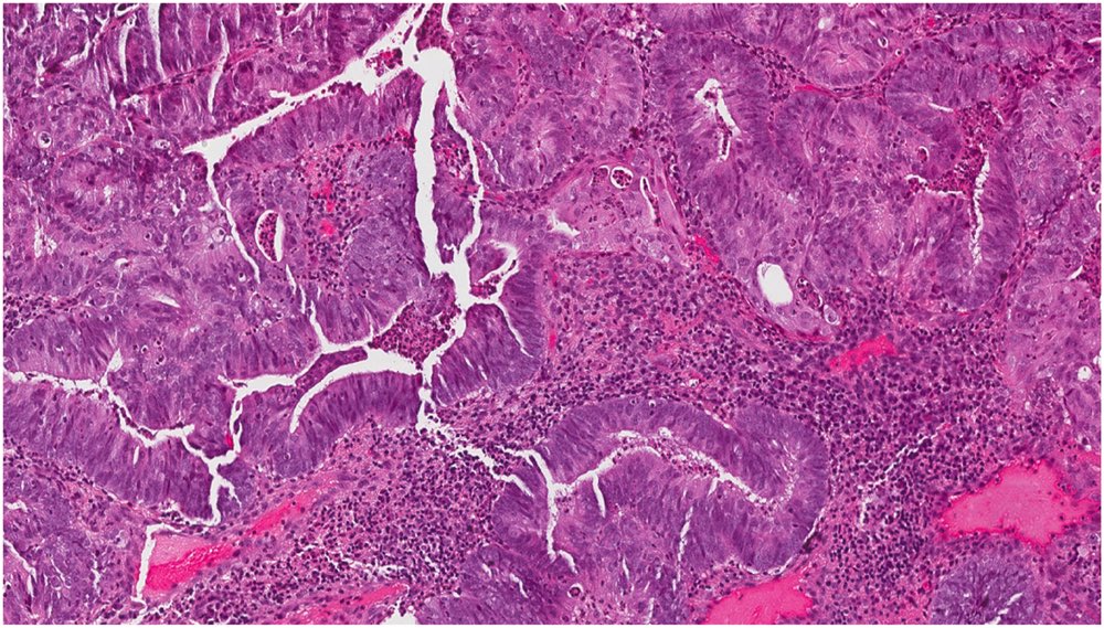
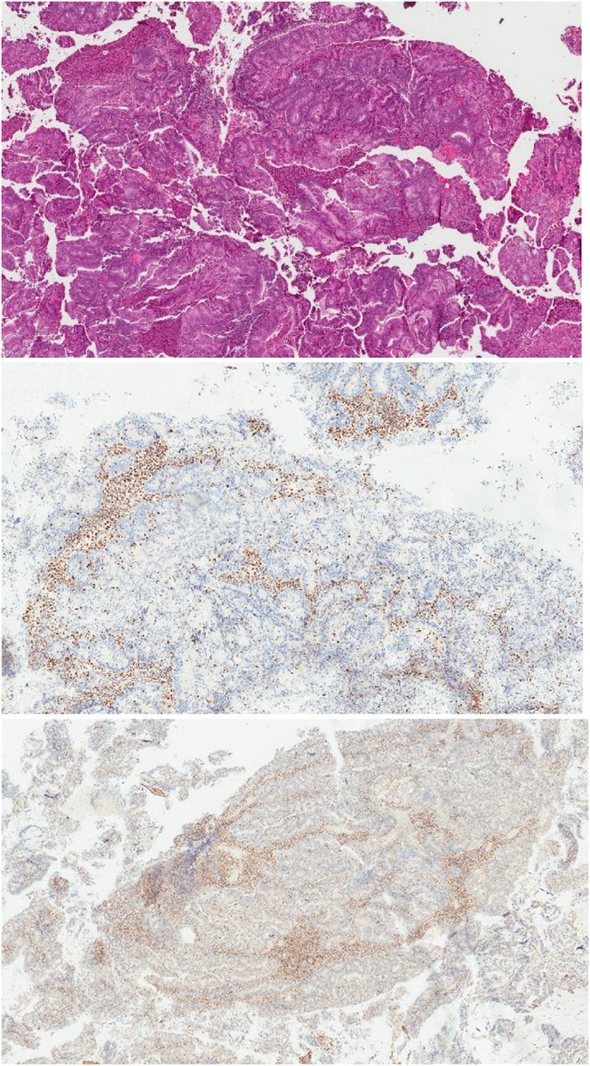
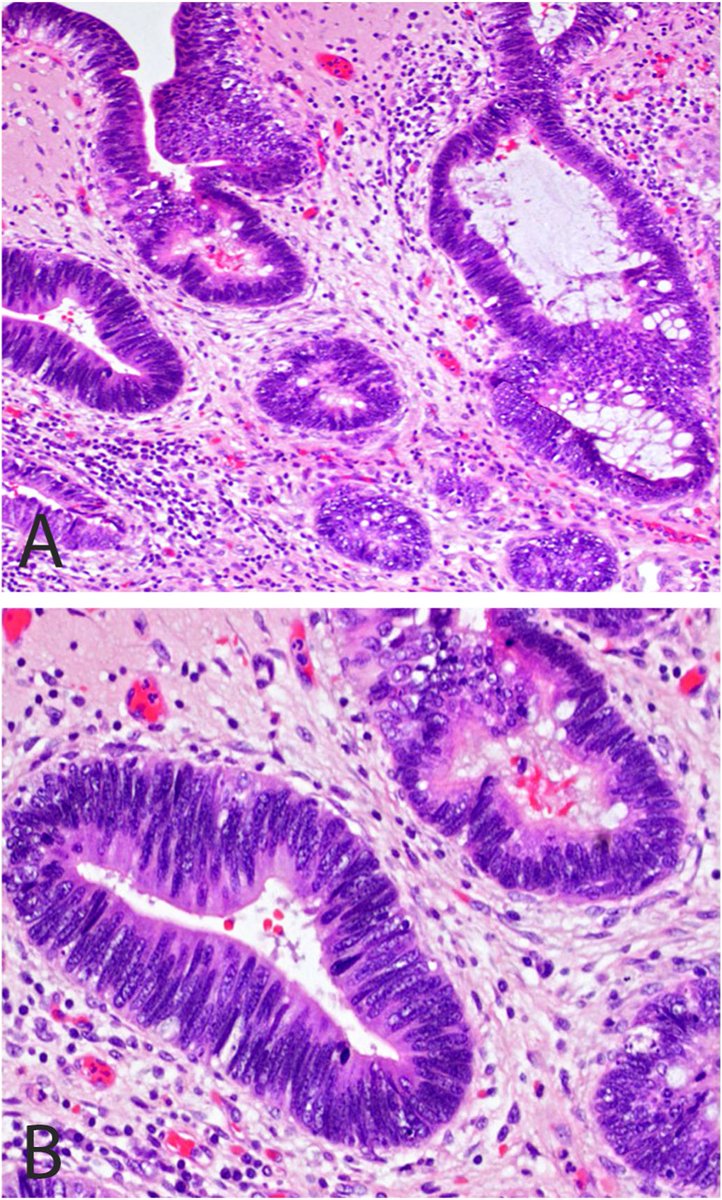
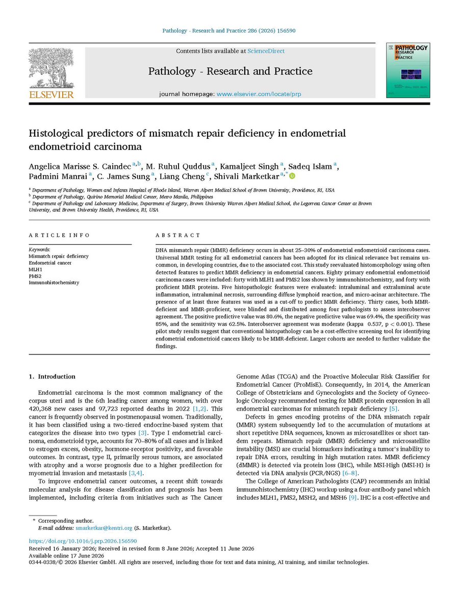
What Histologic Features Predict Mismatch Repair Deficiency in Endometrial Endometrioid Carcinoma?
Delighted to share this article, recently published in Pathology - Research & Practice, led by Drs. Marketkar and Caindec @BrownPathology. Our investigation demonstrates that routine histopathologic evaluation can help identify tumors likely to harbor MLH1/PMS2 deficiency. Five morphologic features were evaluated: microacinar architecture, intraluminal acute inflammation, intraluminal necrosis, extraluminal acute inflammation, and a dense peritumoral lymphoid response. The presence of three or more of these features predicted MMR deficiency with an 80.6% positive predictive value and 85% specificity, with moderate interobserver agreement. These findings support a practical, cost-effective approach for triaging cases for MMR testing, particularly in resource-limited settings where universal testing may not be feasible.
Honored to be part of this study!
Free full-text access:
https://t.co/Z6ZyWfwhwu
Journal article:
https://t.co/k6ziR1qCvw
🌍 Join leading international experts for the SEAUPS–ISUP Webinar! We look forward to seeing you online!
📅 25 Aug 2026 via Zoom 🕗 20:00 (UTC+7) | 06:00 (UTC−7)
Register: https://t.co/UJeieJVKQr
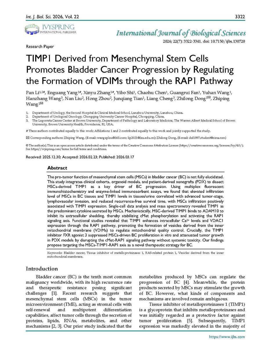
What is the role of mesenchymal stem cells in bladder cancer progression and the evolving landscape of targeted therapy and precision oncology?
Delighted to share this article published this month in the International Journal of Biological Sciences (Journal Impact Factor: 11.7). In this study, Dr. Zhiping Wang and colleagues identify mesenchymal stem cell (MSC)-derived TIMP1 as a critical driver of bladder cancer progression. Using clinical specimens, single-cell transcriptomics, organoid models, and patient-derived xenografts, the authors demonstrate that increased MSC infiltration and elevated TIMP1 levels are associated with advanced stage, lymphovascular invasion, and poorer clinical outcomes. Mechanistically, MSC-derived TIMP1 activates the cMet–RAP1 signaling pathway, promoting mitochondrial quality control through the formation of vesicles derived from the inner mitochondrial membrane (VDIMs), thereby enhancing tumor growth.
Importantly, pharmacologic inhibition of TIMP1 signaling suppressed tumor progression in preclinical models without significant toxicity, highlighting the MSC–TIMP1–RAP1 axis as a promising therapeutic target and opening new avenues for precision oncology in bladder cancer.
Great honor to be part of this important study!
PubMed: https://t.co/XeviHKsyvS
Can you predict FGFR3 mutations in #BladderCancer based on morphology? I am pleased to share this article by Dr. Katrina Collins @katcollmd , which will appear in the August 2026 issue of Laboratory Investigation @LIjournal@virchow
In this study, we found FGFR3 alterations in 17% of urothelial carcinomas, with S249C and Y373C accounting for most mutations. Remarkably, 76% of FGFR3-altered tumors exhibited variant histology, most commonly micropapillary (39%) and squamous differentiation (21%). Both morphologic patterns were exclusively associated with the canonical S249C or Y373C hotspot mutations. Although morphology cannot replace molecular testing, recognizing these histologic patterns may help identify patients most likely to benefit from FGFR3 testing and targeted therapy, particularly in advanced urothelial carcinoma.
Free to download: https://t.co/3fn5v9jF8N
🚨 NEW IN @EUplatinum! 🚨
The largest analysis of focal therapy by Imperial Prostate and UK colleagues, reporting 1⃣0⃣-year outcomes after focal HIFU and cryotherapy in 3⃣4⃣7⃣7⃣ men 🔥🧊
Led by @AJWLight@LondonProstate1@TaimurShah
Read for free: https://t.co/BL19aINTgM
Share your input — help shape the discussion at the ISUP Multidisciplinary Expert Consultation Conference on Precision Biomarker Testing by completing our brief global pre-meeting survey by July 31, 2026.
🔗 https://t.co/LxAbkmMm4g
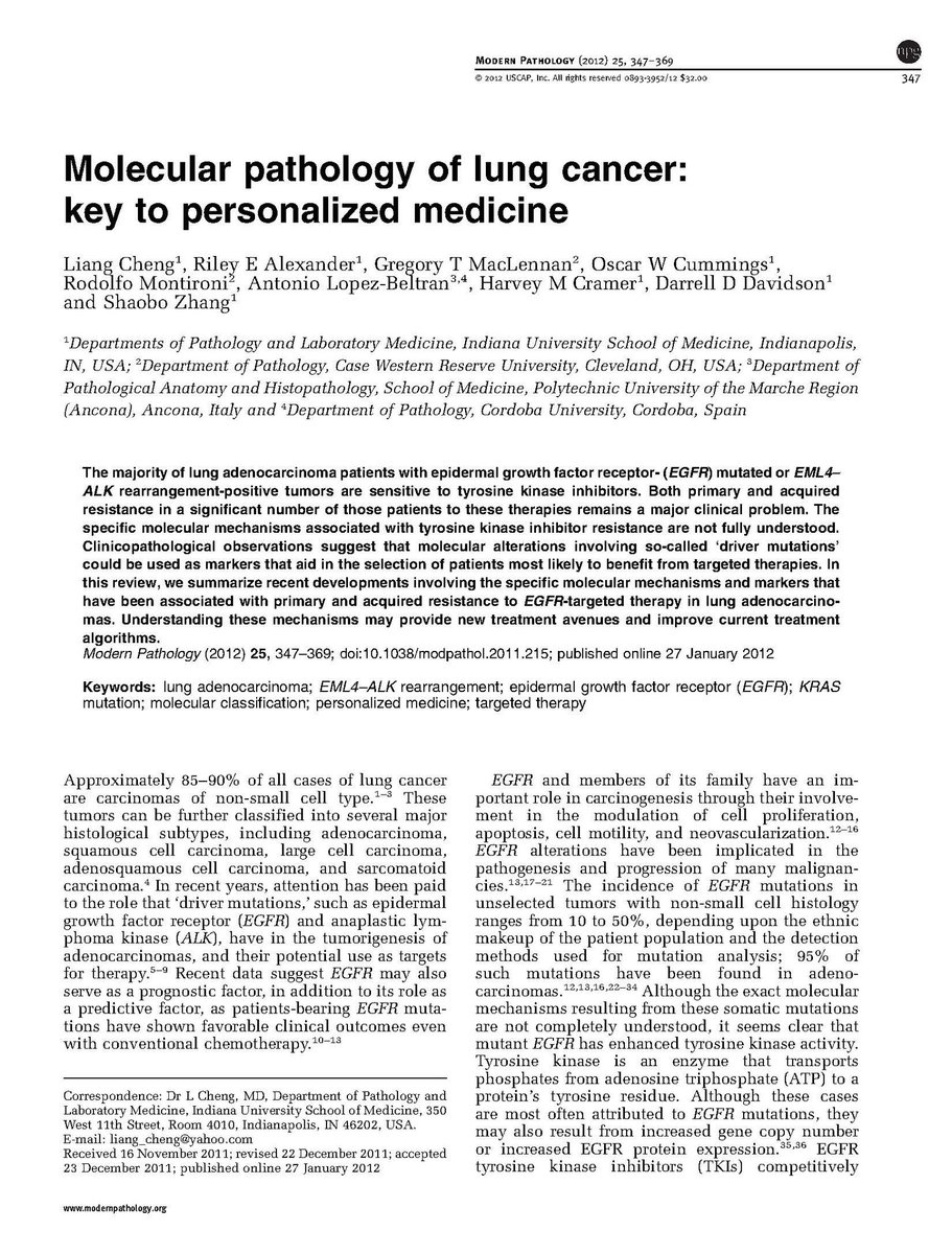
Upon reflection, it is interesting to look back at this comprehensive 23-page review on the molecular pathology of lung cancer that we published @ModernPathology more than a decade ago. At the time, the concepts of driver mutations, molecular classification, and personalized therapy were just beginning to transform the management of lung and other cancers. We summarized the evidence supporting molecular testing in routine surgical pathology practice and proposed practical algorithms for integrating genomic biomarkers into routine clinical care.
I am delighted that this article has now been cited more than 330 times. It has been incredibly rewarding to witness how far precision oncology has advanced over the past decade. Lung cancer has unquestionably led this revolution, demonstrating how molecular diagnostics can fundamentally reshape tumor classification, therapeutic decision-making, and patient outcomes.
Around the same time, our group also published an important study in the Journal of the National Cancer Institute demonstrating that the majority of multifocal lung cancers share a common clonal origin, providing molecular evidence that many multifocal tumors represent intrapulmonary metastases rather than independent primary tumors. This work had important implications for the staging and clinical management of patients with multifocal lung cancer.
Today, many other tumor types - including bladder and prostate cancer - are rapidly following a similar trajectory. The integration of molecular pathology, targeted therapies, liquid biopsy, artificial intelligence, and biomarker-driven clinical trials is transforming oncology and pathology practice across virtually every organ system. What began in lung cancer has become the blueprint for precision oncology throughout modern medicine.
It has been a privilege to witness - and contribute to - this extraordinary transformation. The future of cancer diagnosis and treatment has never been more exciting!
Modern Pathology article: https://t.co/nJn2Em3m6P
JNCI clonal origin study: https://t.co/mf3GyV4te8
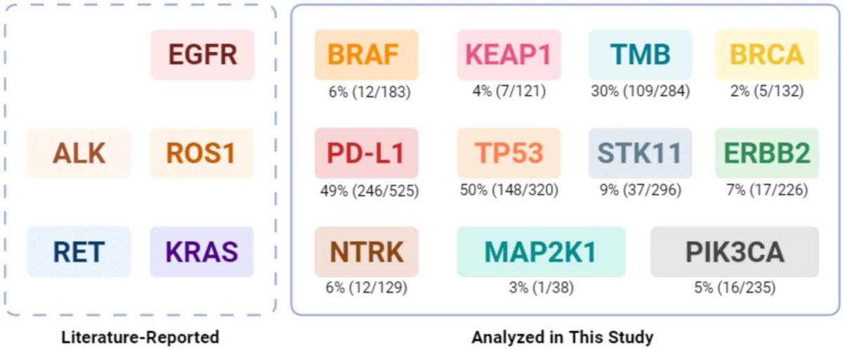
What’s Genomic Drivers of Brain Metastases in Lung Cancer? I am delighted to share our article published in Neuro Oncology Advances @EditorNeuro by Dr. Wang and her colleagues Drs Liu and @drabbaseabbas@bcarneiro7@mhadfield30@BrownPathology@BrownUCancer
Brain metastases remain one of the most devastating complications of advanced non-small cell lung cancer (NSCLC), affecting up to half of patients during the course of their disease and significantly compromising both survival and quality of life. In this comprehensive systematic review and meta-analysis of 19 studies involving 3,028 patients, we provide an updated synthesis of the genomic landscape driving brain metastases and its implications for precision oncology.
We identified TP53 mutations (50%), PD-L1 overexpression (49%), and high tumor mutational burden (30%) as the most prevalent molecular alterations associated with brain metastases. Additional clinically actionable but less frequent alterations included ERBB2, BRAF, NTRK, PIK3CA, STK11, and KEAP1, highlighting the remarkable molecular heterogeneity of metastatic NSCLC and the expanding opportunities for targeted therapies and immunotherapy.
Importantly, our study demonstrates substantial genomic discordance between primary lung tumors and matched brain metastases, underscoring the importance of site-specific molecular profiling to guide individualized treatment decisions. The authors also review emerging targeted therapies, immunotherapeutic strategies, and ongoing clinical trials, providing a timely roadmap for integrating biomarker-driven precision medicine into the management of patients with NSCLC and brain metastases. This outstanding review will serve as a valuable resource for oncologists, pathologists, translational researchers, and neuroscientists seeking to improve outcomes for patients with this challenging disease
Full article: https://t.co/oBfq3Rhnmp
https://t.co/oyQaAWrTgU
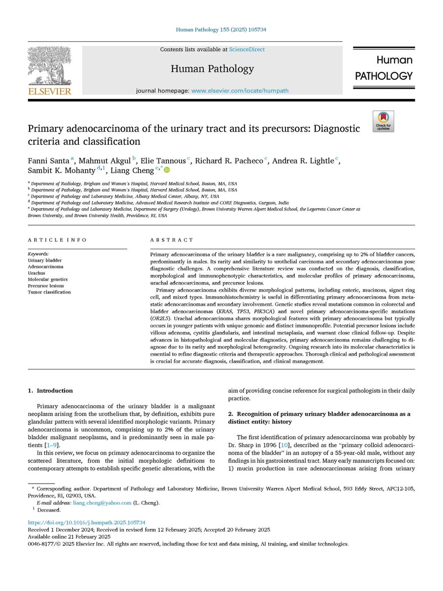
Comprehensive review on diagnostic criteria and classification of malignant glandular lesions of the urinary bladder by Dr. Fanni Santa @FV_Santa and her colleagues, published as the featured cover article @Human_Pathology.
This review provides a practical framework for one of the most diagnostically challenging areas in genitourinary pathology, integrating contemporary morphologic, immunohistochemical, and molecular advances into a unified diagnostic approach.
Dr. Fanni Santa and her colleagues address the classification and differential diagnosis of primary bladder adenocarcinoma, urachal adenocarcinoma, Müllerian-derived tumors, and precursor lesions, emphasizing key diagnostic pitfalls in distinguishing these uncommon neoplasms from more common primary bladder tumors and secondary involvement by tumors from other organs. The review also highlights emerging molecular insights, including recurrent genomic alterations with potential therapeutic implications, and discusses evolving concepts surrounding precursor lesions such as villous adenoma, cystitis glandularis with intestinal metaplasia, and adenocarcinoma in situ.
This comprehensive review is an essential resource for pathologists, urologists, oncologists, and trainees, and hope it will contribute to improved diagnostic accuracy, multidisciplinary communication, and optimal patient management for these rare but clinically significant tumors.
Read the full article here:
https://t.co/hHpwZz0VKM
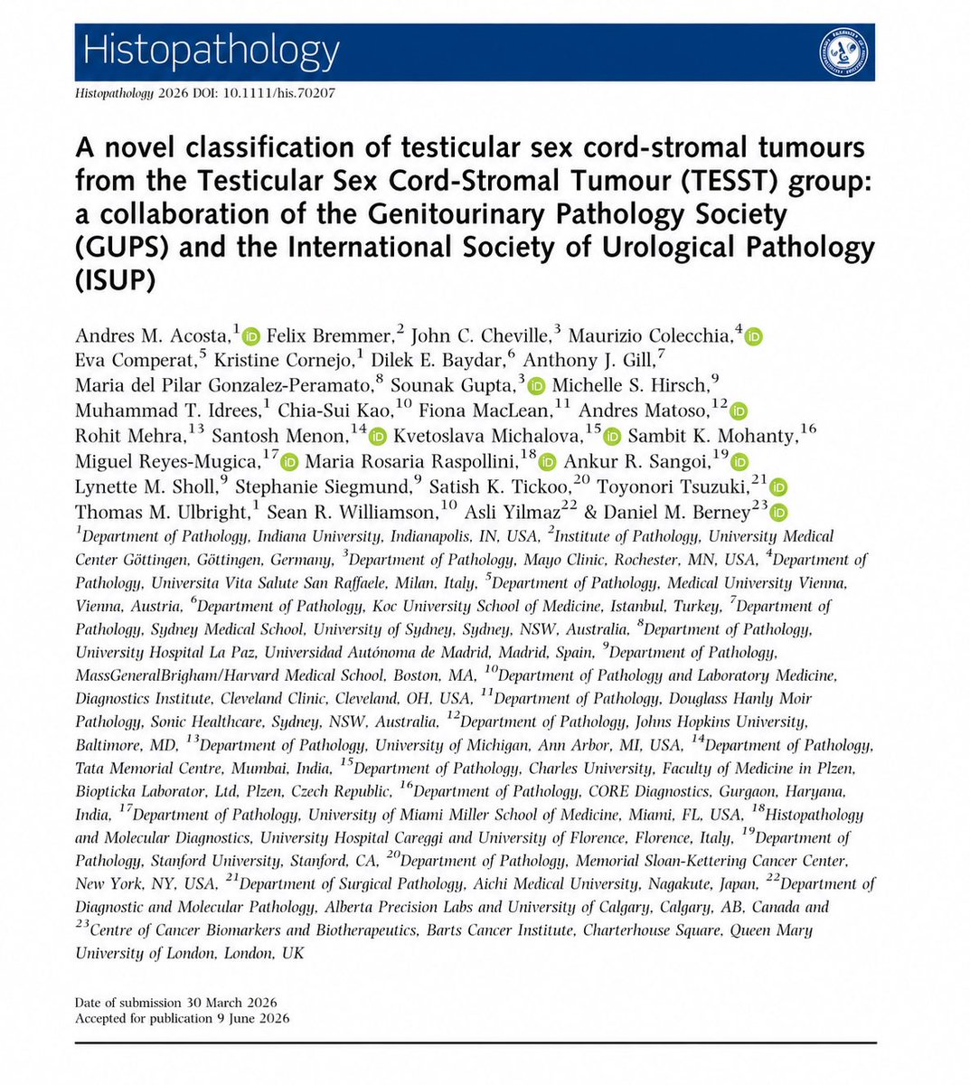
New in @Histo_Journal: Collaboration with @GU_Path_Society and @IntSocUropath, introduces a novel classification of testicular sex cord-stromal tumors, integrating morphology and molecular pathology to improve diagnostic consistency #openaccess
🔗 https://t.co/bp9K00Ig8f
Good news to share! Our paper, "Hereditary Renal Cancer Syndromes: Redefining Clinical Management in the Era of Precision Medicine", has been accepted for publication in European Urology (journal impact factor:29)!
It has been a true honor and privilege to work with Dr. Fanni Santa @FV_Santa , an exceptionally talented and dedicated physician-scientist. Fanni completed this work during her observership at @BrownPathology last month - an extraordinary achievement that speaks to her intellectual curiosity, work ethic, and passion for academic medicine. Fanni is undoubtedly one of the brightest rising stars in genitourinary pathology, and I look forward to many more collaborations and to watching your continued success.
⭐️Nice review in @JAMAOnc 👉 Circulating Tumor DNA in Early Breast Cancer.
👉Multiple ongoing prospective interventional trials are evaluating whether ctDNA-guided treatment escalation or de-escalation can improve patient outcomes and support the routine implementation of ctDNA assays in clinical practice https://t.co/SGVdgJhk9x
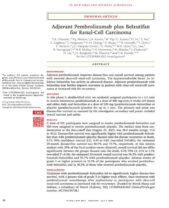
🚨 Results from #LITESPARK022 (#LS022) are out in @NEJM!
1/This phase III trial evaluated whether adding the HIF-2α inhibitor Belzutifan (BEL) to adjuvant pembrolizumab (PEMBRO) could improve outcomes for patients with resected clear-cell RCC at increased risk for recurrence.
https://t.co/1BLAercj3p
@oncoalert@asco@myESMO