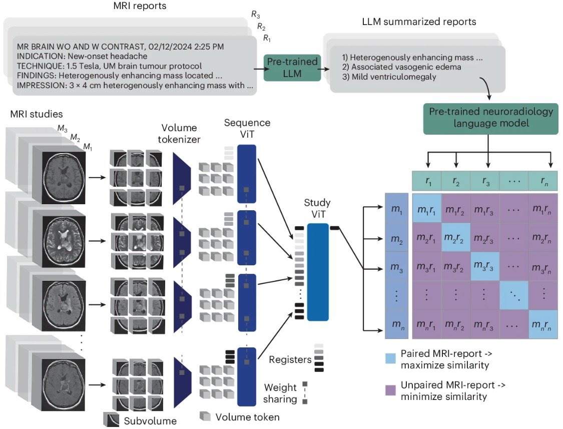
We present Prima, the first medical foundation model for neuroimaging that supports full real-world, clinical MRI studies: the whole MRI in >>> diagnosis out!
Thanks to @umichmedicine, @umichneuro, and our @mlins_lab.
https://t.co/VKZBqtbZPW
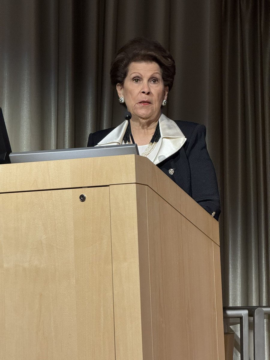
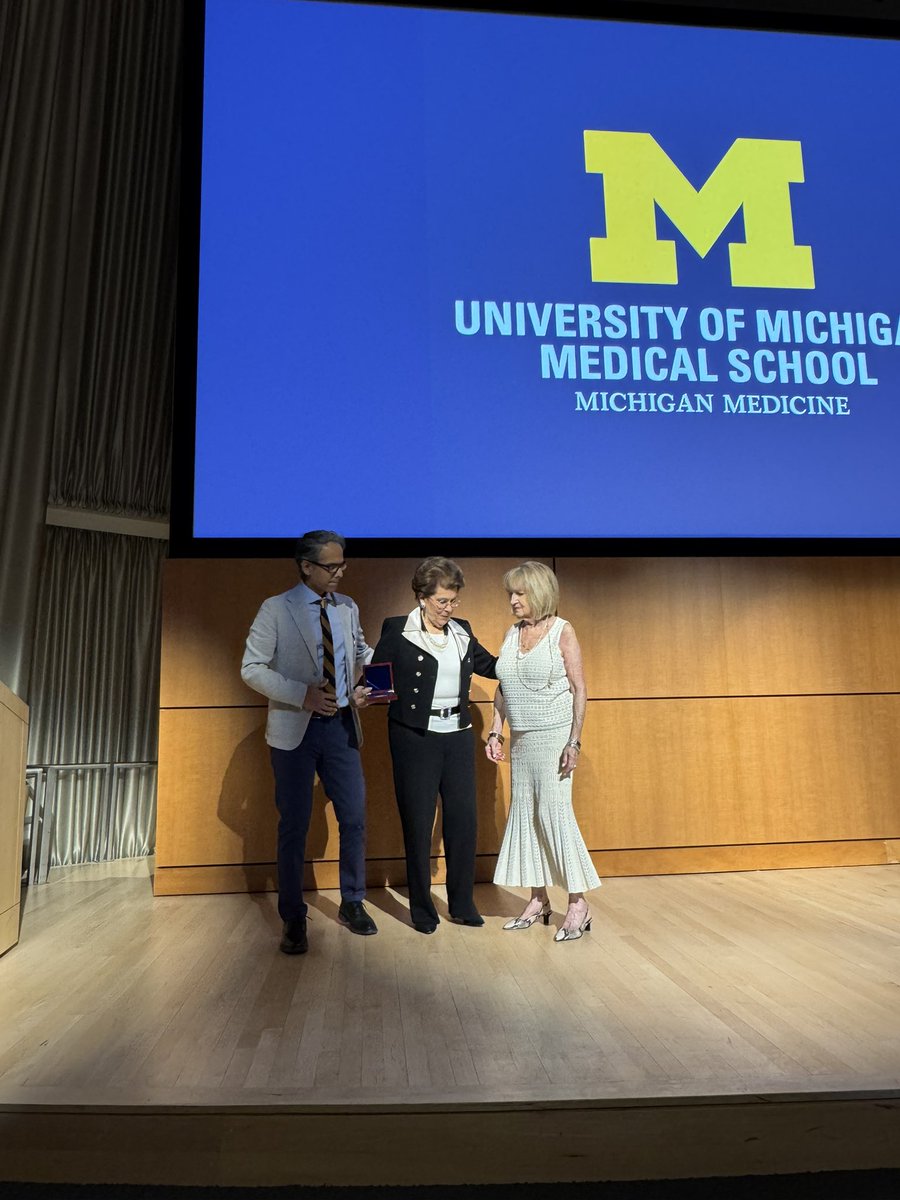
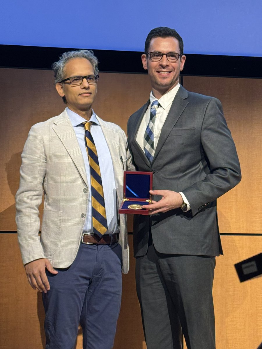
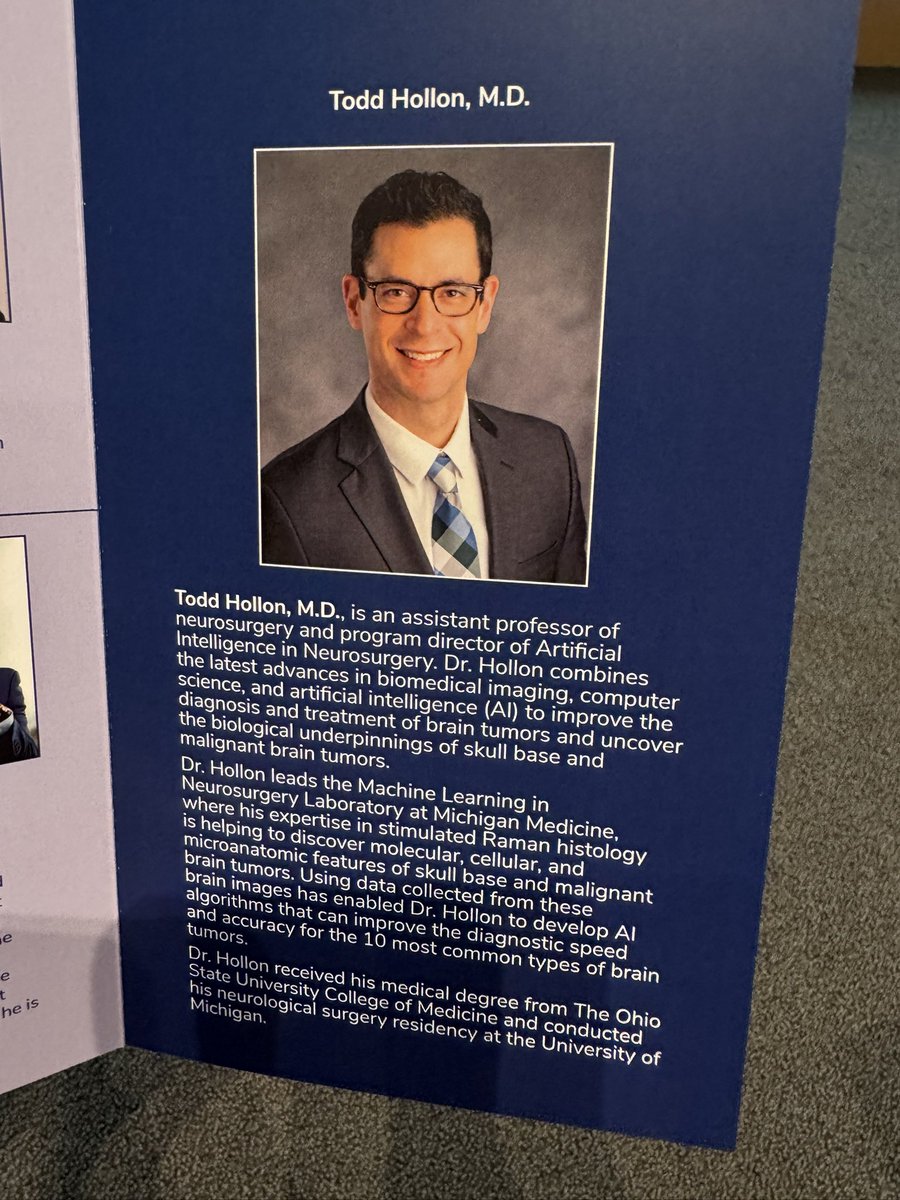
A well-deserved congratulations to @ToddCHollon for being honored as the inaugural holder of the Joseph R. Novello, MD and Alfredo Quiñones-Hinojosa, MD Research Professorship!
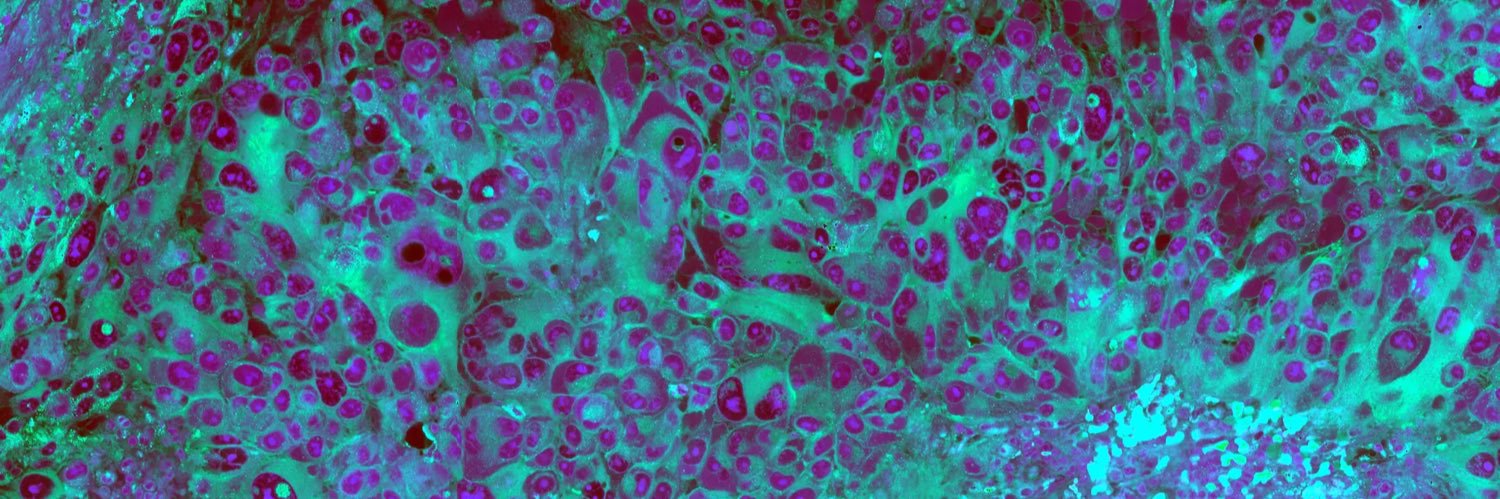
Real-time intraoperative brain surgery with a visual foundation #AI model:
In less than 10 seconds, accurate detection of tumor (glioma) infiltration from fresh surgical tissue without labels, validated in a prospective multi-center trial, made open-source @Nature@ToddCHollon@umichneuro @HerveyJumper @avkondepudi and colleagues
https://t.co/tnV1hx4WMB
🚀 Proud to introduce #FastGlioma: the first foundation model enabling rapid, accurate detection of brain tumor infiltration during surgery, in under 10 seconds. With FastGlioma, we’re minimizing the risk of residual tumor and enhancing outcomes for glioma patients. This work sets a new standard in real-time, microscopic-level detection, powered by AI in healthcare. Kudos to the incredible team @mlins, @HerveyJumper, @DanOrringerMD, @InvenioImaging, @CameloPiragua!
Read the full paper in @Nature: https://t.co/gJ3dCPYod4
4/7 📊 Hou & Jiang et al. present SPT, a framework for learning self-supervised slide representations, which is consistently able to learn strong slide-level features across a variety of encoders, including UNI. arXiv: https://t.co/2FEQ9Kcaau
⚡️This is the first work to investigate slide pretraining across a diverse variety of ROI encoders. The analyses in Hou & Jiang et al. suggest that slide pretraining provide the biggest performance gains in less powerful ROI encoders, with least benefit in HiDisc and UNI. They also show the importance of further finetuning as well, which can yield as big of an improvement as slide pretraining.
💭 We believe more development needs to happen in self-supervised slide encoders than ROI encoders. Few works in this area, and where most of the technical advancements need to be made 🔥
🎉 Thanks to all who joined us for a #CVPR2024 Michigan AI meetup, it was fantastic connecting with everyone!
Looking forward to more gatherings like these!
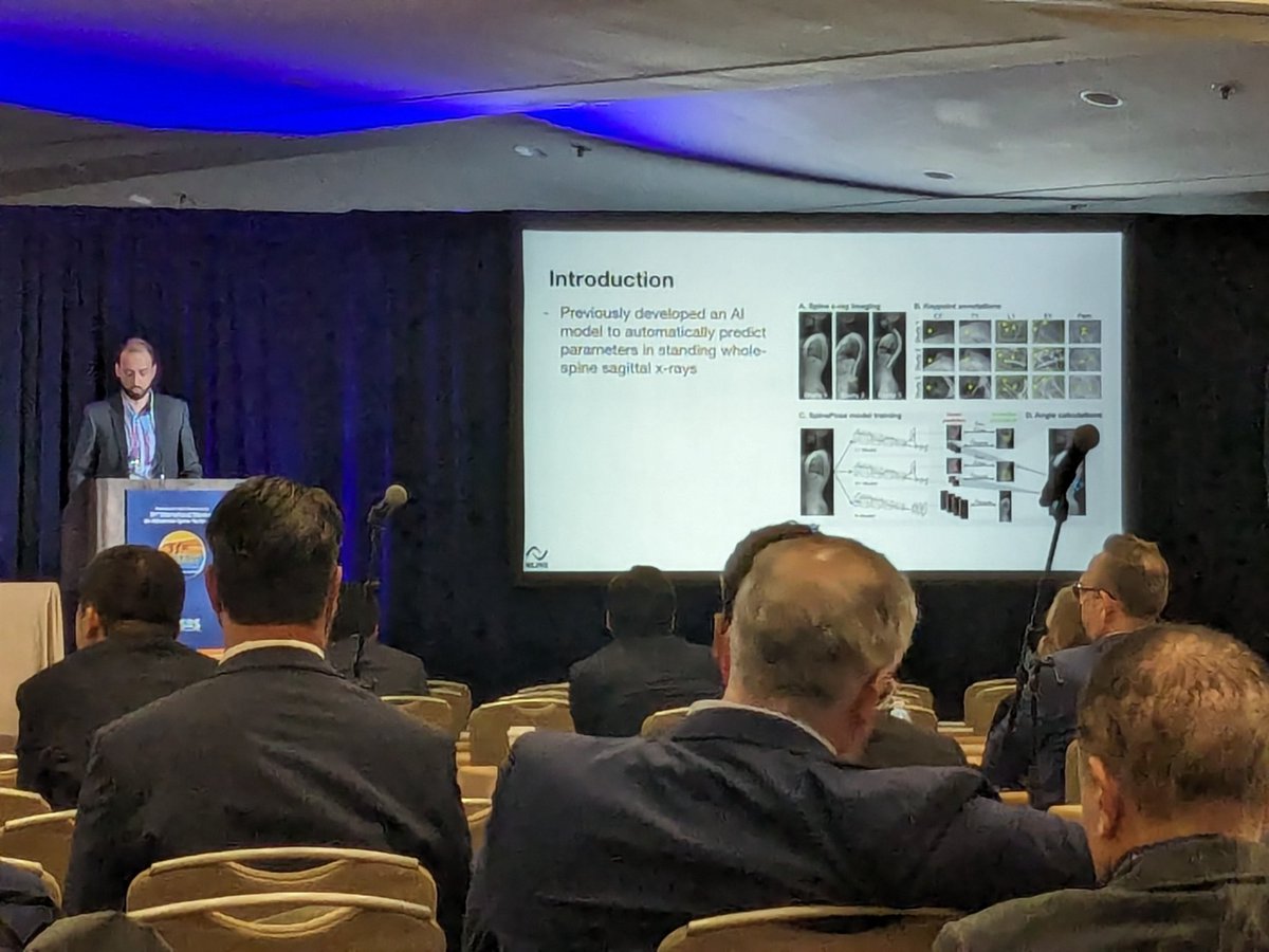
Very excited to share our most recent work out of the @mlins_lab - SpinePose is a novel deep learning model that predicts spinopelvic parameters. Special thanks to @ToddCHollon , Dr. Paul Park, @joseph_linzey@__chengjia__ https://t.co/t2DQifUjY3
Great work from our @SamirHarake and @mlins_lab! Using AI to make spine surgeon's lives just a bit easier.
Development and validation of an artificial intelligence model to accurately predict spinopelvic parameters https://t.co/KmNUQyFO1J
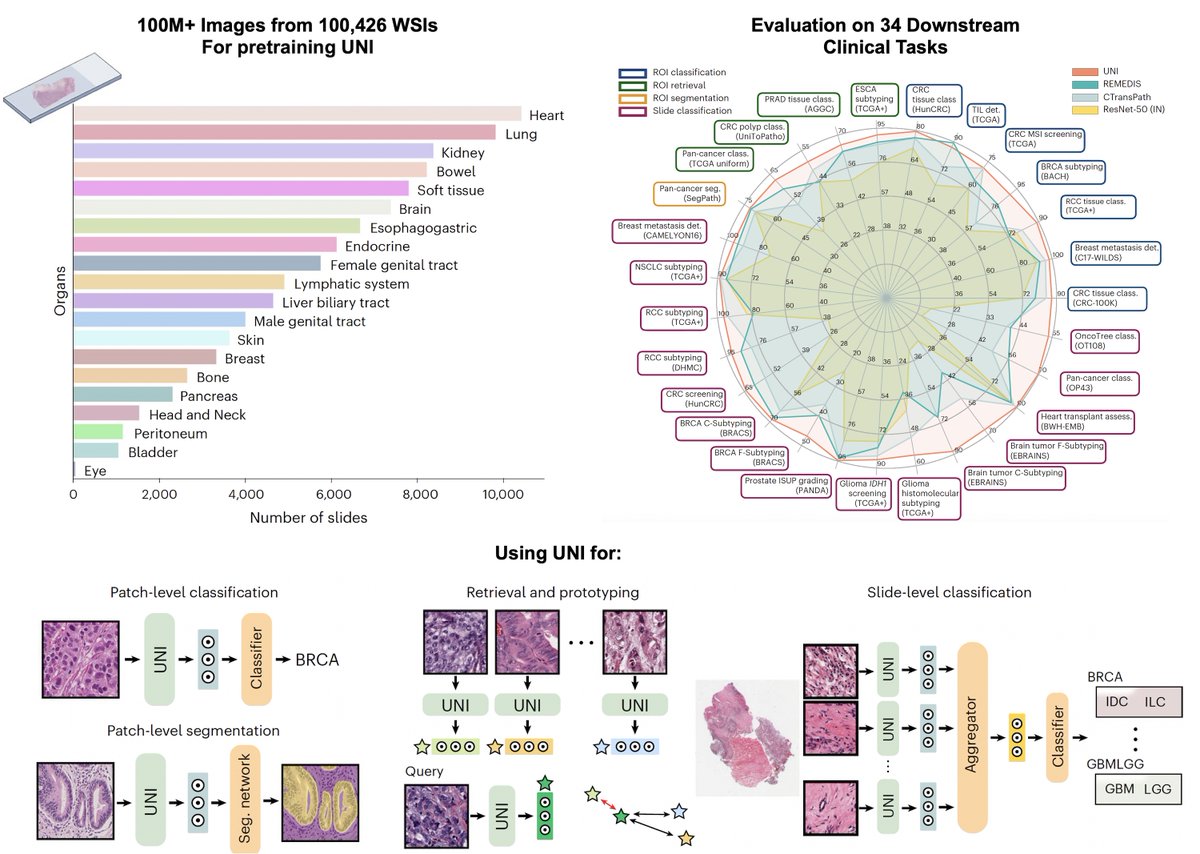
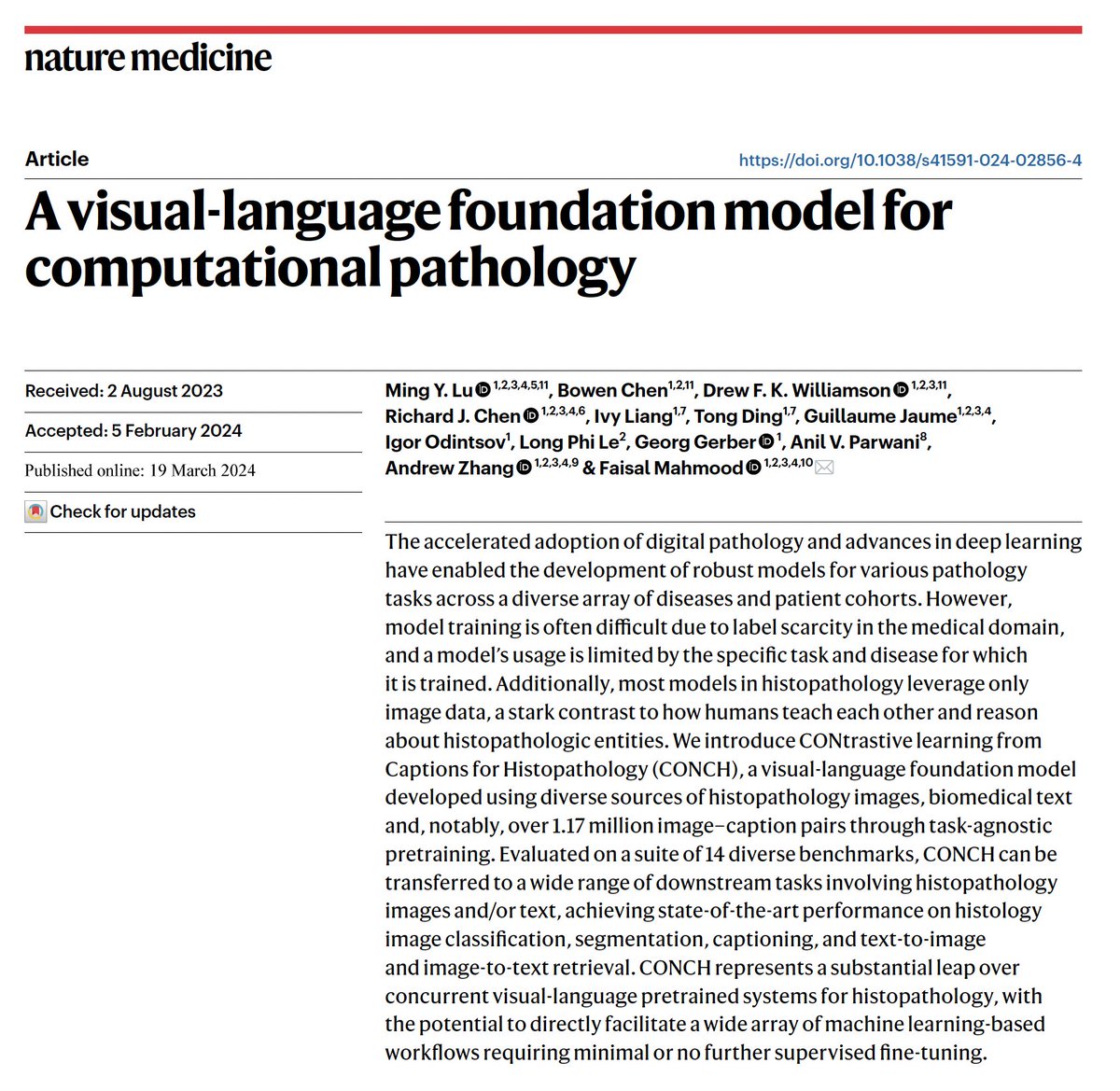
⚡️🔬📣Excited to share our two new @NatureMedicine articles, we develop computational pathology foundation models,
1. UNI, a self-supervised computational pathology model trained on 100 million pathology images from 100k+ slides.
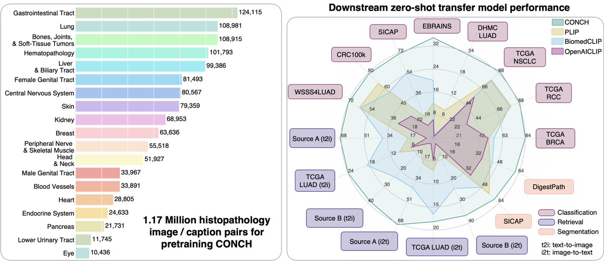
2. CONCH, a vision-language model for computational pathology trained on 1.17 million pathology image-text pairs.
Access the articles @NatureMedicine
UNI: https://t.co/f207RP0JKs
CONCH: https://t.co/9eHXwjZMub
Access the code, models:
UNI: https://t.co/5Gkyzd8R8a
CONCH: https://t.co/BLG2G3bTuO
Interesting aspects:
- Both models are evaluated on a host of different clinically relevant tasks for WSI classification, ROI classification, segmentation, image retrieval, image-to-text retrieval, text-to-image retrieval, in 0-shot, few-shot and supervised settings. These adaptations encompass the utility of large public datasets and evaluations on independent test cohorts.
- Both models exclude commonly used public computational pathology benchmarks from pre-training allowing for a much more holistic evaluation.
Some limitations: Both UNI and CONCH represent early developments in foundation models for pathology. More data, and additional evaluation is needed to realize the full potential of these models. Nevertheless, we show the models capabilities on a variety of different benchmarks with several demonstrating state-of-the-art performance.
Future work and insights: While these developments are exciting, they represent work we did about a year ago when the pre-prints were made available, since then we have been busy collecting significantly larger datasets and hope to make larger models available in the future. We have also used UNI and CONCH as the backbone for our Pathology specific chatbot, PathChat (https://t.co/OuVsJvrLTQ), which is further trained on hundreds of thousands of pathology specific Q-A instructions.
We are also excited to see foundation models for several other areas of biomedicine including for single cell data (https://t.co/vkvE3ulri9), radiology (https://t.co/c5CLbgmcrG) and the general trajectory towards general purpose AI for biomedicine.
Congratulations to our superstar leaders @richardjchen@MYLu97 @DFKW_MD @TongDing99, Bowen Chen and everyone else who contributed to these studies @GuillaumeJaume@GreatAndrew90@sharifa_sahai@Aparwani_dpath and others.
Our @SamirHarake won the Kuntz Scholar Award at @spinesection. Not bad for his first deep learning paper.
Check out the project website!
https://t.co/H6xWPvKCAd
Our @__chengjia__ has just received his NIH F31 from @NIH_NINDS on using optical imaging and deep learning for label-free single-cell phenotyping. Looking forward to some impressive work over the next two years!