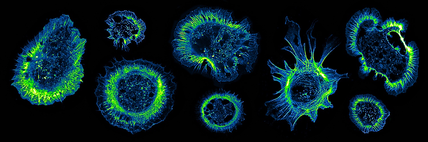
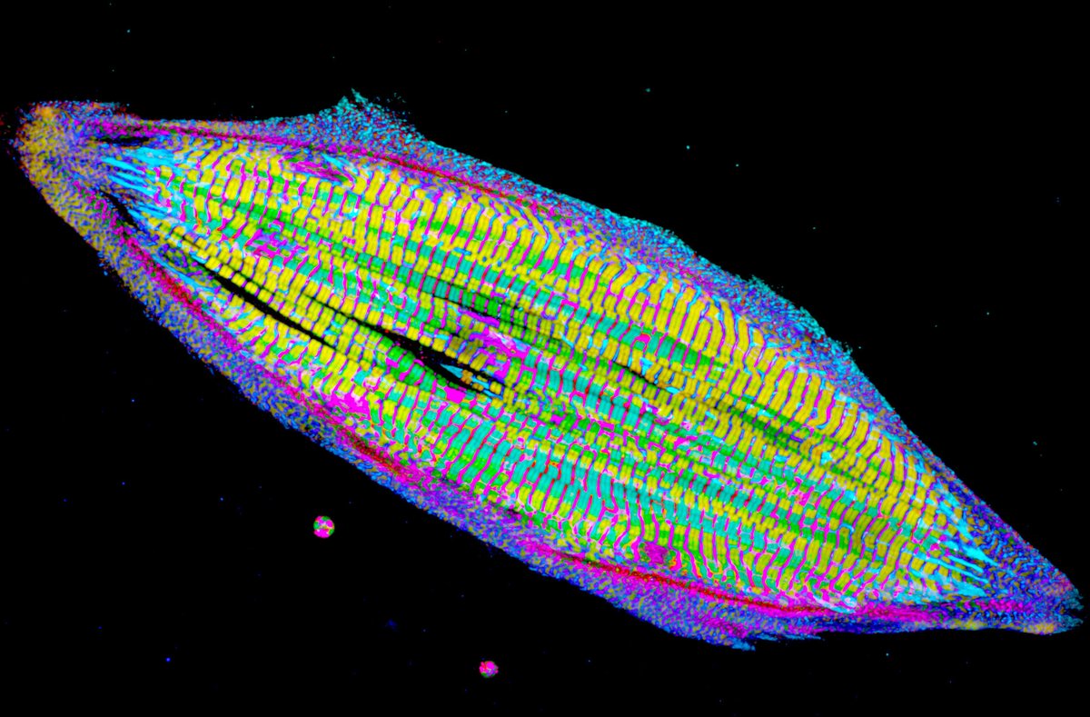
Imaging-based CRISPR genome screening allows effects of cell-regulating molecules & modifications to be revealed in live & fixed cells – applied here to frog embryo cilia
📹 Jingbo Sun et al @DavidBreslowLab@Yale in @Dev_Cell
➡️ https://t.co/DAPis1jjfe
An iPSC-cardiac myocyte (heart muscle cell) surrounded by cardiac myocyte-derived small extracellular vesicles (tiny dots; AKA cardiosomes) and two cardiac myocyte-derived large extracellular vesicles (thankfully just called that; I love descriptive names). #CellBiology
A new MRI protocol described in @ScienceAdvances can scan the human #brain and its venous network in under 7 minutes, marking it as a potential tool for diagnosing cerebrovascular diseases and monitoring neurodegeneration. https://t.co/5tiRCjdpd0
I’m absolutely thrilled to share this! 💥
👉 A breathtaking super-resolution image reveals interdigitated podocyte foot processes (magenta) and the slit membrane (green) within a glomerulus.
The third stain (blue) indicates Claudin-5 expression, a tight junction protein and biomarker that helps identify which podocyte foot processes—still narrow and healthy—are already restructuring and which are likely to become effaced later. 🚀
In the top image (higher magnification), an intermediate podocyte foot process (iFP) shows a distinct linear blue signal (star) instead of a missing or punctate pattern (arrow).
In contrast, the lower image displays an elongated Claudin-5 line in an effaced foot process (eFP
This opens up an exciting opportunity—not only to get more information from kidney sections, which usually represent just static snapshots—but also to gain insight into potential future structural changes 👏.
👉 More on this in my presentation at this year’s ASN Meeting in November at Houston.
I hope to see you there!
Amazing work of @NIPOKA_Podocyte & Universitätsmedizin Greifswald 👏
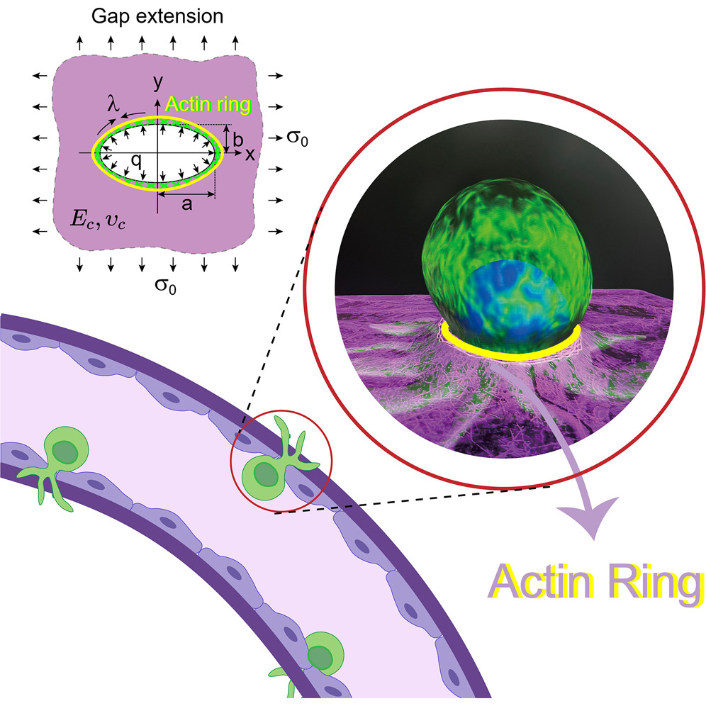
🧪 Through experimental approaches and theoretical modeling, scientists showed that the actin ring contraction generates significant mechanical resistance, impeding tumor cell migration and inducing tumor cell apoptosis.
https://t.co/f6sRh5U0ov
The two aggregates immediately fused and began to self-organize as a single structure. Anterior (WNT-free) cells intercalated to the center of the posterior aggregate to form hindbrain, a continuous neural tube, and bilaterally segmented somites. (4/n)
For this #FluorescenceFriday, an image of an adult mouse kidney labeled with AQP2 and alpha SMA antibodies. AQP2 (green) marks the collecting duct and distal connecting segment while SMA marks the arterial tree. Courtesy of talented postdoc Sarah McLarnon.
How do we measure the interaction between astrocytes and vessels?
We 3D reconstruct how astrocytes enwrap the vessels depending on the conditions we have.
For #FluorescenceFriday you have the image on the left and the reconstruction on the right. Red =GFAP, pink = vessel (CD31)
A single aquaporin protein lets one billion water molecules into a cell each second. It is one of the "fastest" channels in all of biology.
Each water flows through in single file, while H+, sodium, potassium, and other ions are excluded.
What explains this remarkable selectivity? Two clever protein filters.
But first: Each aquaporin protein is made from six alpha-helices that run back and forth through the cell membrane. These helices form an hourglass shape so that the channel is cone-shaped at either end and extremely narrow in the middle.
The two ends of the protein also mirror each other, with their point of symmetry occurring at the narrowest part of the channel. It's thought that modern aquaporins evolved through a gene duplication event.
Now let's zoom in a bit and look at the two filters within aquaporin that account for its extreme selectivity. Water can flow either way through the channel, so we'll imagine that we're moving from OUTSIDE the cell to INSIDE the cell.
The first filter we encounter is called the ar/R filter. This is a cluster of polar and charged amino acids that point inside the aquaporin channel.
These four amino acids are *usually* arginine (+ charge), histidine (polar), and two bulky amino acids like phenylalanine and tryptophan.
These four amino acids come together to restrict the middle of the pore, making it just wide enough for a single water molecule to pass. The positively charged arginine also helps "stabilize" water molecules while forcing away sodium ions or protons.
The second filter encountered on our journey into the cell is called the NPA motif (short for asparagine–proline–alanine). There are two of them inside each aquaporin.
These two protein motifs point at each other in the center of the aquaporin. As a water molecule passes the ar/R filter and moves deeper into the channel, it forms hydrogen bonds with the asparagine amino acid in the first NPA motif.
But because the two motifs are oriented in opposite directions, the local electric field at the channel’s midpoint next forces each water molecule to rotate 180° and latch onto the asparagine in the second NPA motif. In other words, each water molecule gets flipped around inside the aquaporin channel.
This "flipping" is important because it breaks the water molecules free from any hitch-hiking protons. It acts as the final filter.
Zebrafish embryo tail, post amputation, showing cell migration to the wound, cell divisions, and vasculature extension toward the cut. Plasma membranes (green), nuclei (red), and erythrocytes (purple) as seen by adaptive optical lattice light sheet on MOSAIC.
🧬A new imaging technique developed by the Liu Lab uses a novel DNA barcode system to track hundreds of RNA & protein molecules in single cells within thick biological samples, providing a full picture of how these structures are organized inside tissues. https://t.co/GAS8BalQMI
My colleagues put together an ebook on tissue clearing (for #lightsheet microscopy).
If you are like me and have only experience with live imaging and are a bit confused with the different acronyms flying around, I highly recommend you take a look!
I counted 37 methods!
Introducing PetaKit5D: a comprehensive suite of software tools for processing petabyte-scale microscopy data from Janelia Sr Fellow/HHMI Investigator @Eric_Betzig, Xiongtao Ruan, Matthew Mueller, Srigokul Upadhyayula @ABCUCBerkeley@UCBerkeley & colleagues https://t.co/OaVxuj7e0c
I am lucky!
- Spectacular cardioid ♥️ sample from Lavinia Ceci Ginistrelli at IMBA, Vienna @IMBA_Vienna
- Isotropic resolution using AxL Cleared Tissue LightSheet 🔬 @3i_inc
- Breathtaking visualization in syGlass VR 😎
@michael_syglass
Geometric packing of tubules in the developing kidney urinary collecting system leads to tissue stiffening and rhythmic mechanical stresses local to nephron-forming niches that synchronize with tubule branching. @HughesLabPenn
https://t.co/3XEkztIGyM
Super excited to share Bong-Ihn Koh's @bikohcello mind-blowing 🤯🤯🤯 work from @ralfhadams lab, now published in @Nature
Check out the amazing images & tweetorial 👇
https://t.co/eC5sHY1ZuI
https://t.co/aP1i6wQfUN
In recent years we learned how critical the skull bone marrow is as an immune reservoir protecting the brain. Today @Nature, the skull bone marrow is distinct from any others in the body, with lifelong extension, resilient to the aging process (@justsaysinmice )
https://t.co/bxRtGhIwF7
@ralfhadams and colleagues