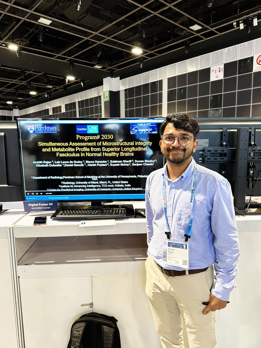
2/2 Key finding: Cho/Cr and Cho/NAA ratios track closely with microstructural metrics (FA, MD, ficvf) along the SLF I & II, with strong reproducibility across scans.
Work with an amazing team @ Penn Radiology, led by @drsuyash & Dr.Sanjeev Chawla
https://t.co/GNiYY6aeKT
2/2 Key finding: Cho/Cr and Cho/NAA ratios track closely with microstructural metrics (FA, MD, ficvf) along the SLF I & II, with strong reproducibility across scans.
Work with an amazing team @ Penn Radiology, led by @drsuyash & Dr.Sanjeev Chawla
https://t.co/GNiYY6aeKT
New preprint 🧠📄
1/2Excited to share our proof-of-concept study fusing whole-brain spectroscopic imaging (WBSI) with diffusion MRI tractometry along the superior longitudinal fasciculus in healthy adults-a step toward a co-localized metabolic + microstructural imaging framework.
Recent advances in targeted therapy are redefining the management of IDH-mutant gliomas.
This review examines the emerging role of advanced imaging biomarkers in detecting IDH mutations and monitoring response to treatment.
Read the full publication: https://t.co/cUrz2rcZM4
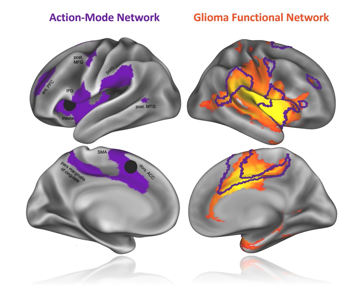
Gliomas do not develop randomly but prefer a specific network: the action mode network -- no matter the tumors are in the cortex or cerebellum. This suggests action-related functional activity may play a key role in tumor growth. @ndosenbach@foxmdphd
https://t.co/Uizts2vuz9
✅✅ A reminder by Dr. Rahaf Ajaj:
She wasn’t on the Stanford list… but she made it to the Nobel stage. 🏅
Mary E. Brunkow, one of this year’s Nobel Prize winners in Medicine, has only 34 published papers and an H-index of 21.
She never appeared in Stanford’s ranking of the world’s top 2% of scientists.
She didn’t chase citations, metrics, or the spotlight.
Yet, she became part of a discovery that changed how humanity understands the immune system.
Today, while many are busy chasing numbers, titles, and rankings —
she reminds us what truly matters in science: the question.
🔹 She wasn’t running after the lists.
🔹 She was running after the truth.
Because in the end, it’s not about how many papers you publish…
It’s about how deeply your idea can reshape the world.
Focus on your idea, not your ranking.
🧠✨ A great overview of tractography—its current state and future directions—by @maxdescoteaux and @tractography!
🎥 Must-watch for all tractographers:
https://t.co/lq2a0l31Pn
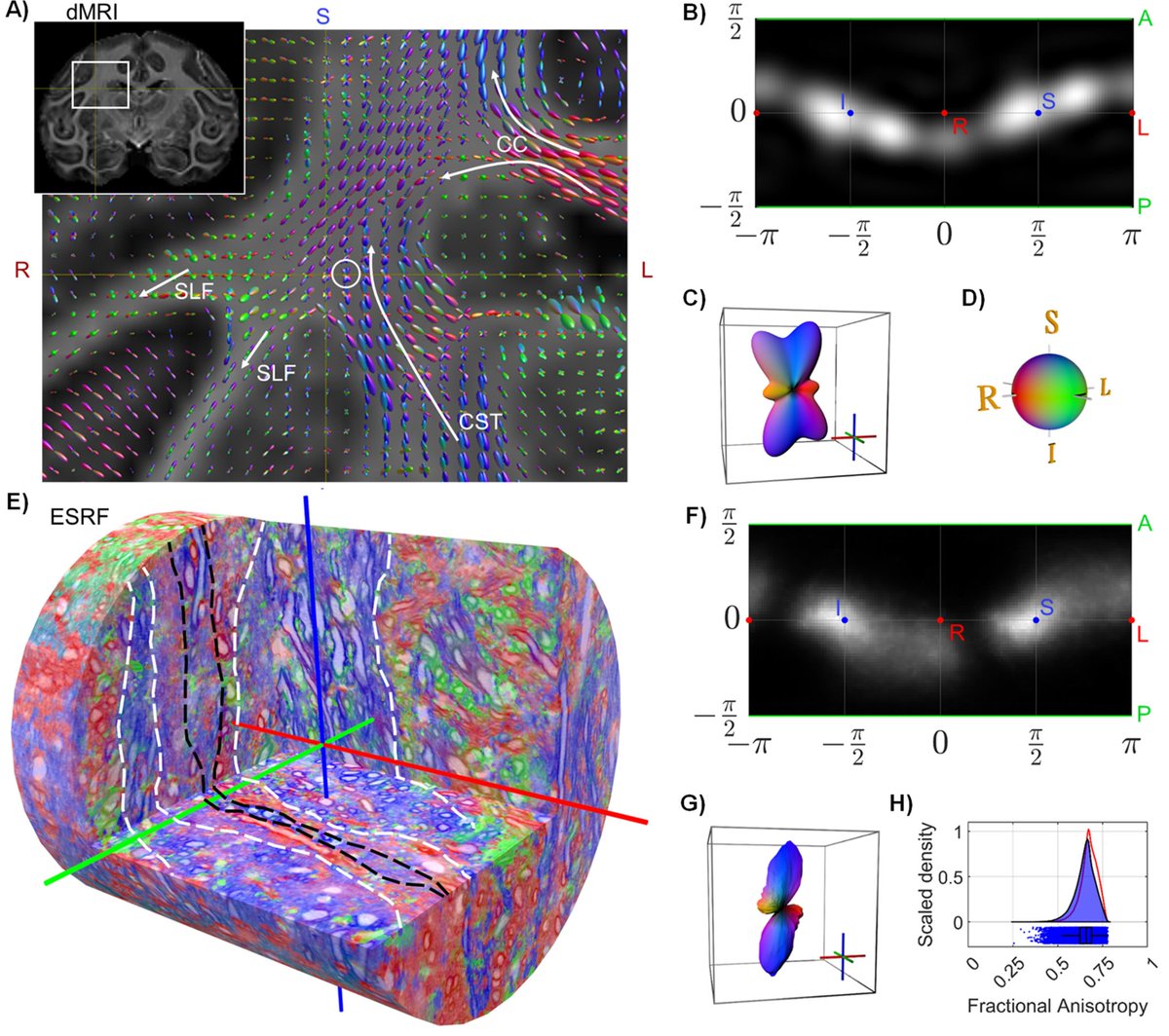
One of the most important recent works in dMRI field by @Tim_Dyrby showing dMRI compared with microstructure.
A lot of insights into brain microstructure and macro structure. 👇🧠
A must read for those working on dMRI.
https://t.co/cZruhAJYG6
Our paper on super resolution for diffusion MRI is finally published
Diffusion magnetic resonance imaging (diffusion MRI) is widely employed to probe the diffusive motion of water molecules within the tissue. Numerous diseases and processes affecting the central nervous system can be detected and monitored via diffusion MRI thanks to its sensitivity to microstructural alterations in tissue. The latter has prompted interest in quantitative mapping of the microstructural parameters, such as the fiber orientation distribution function (fODF), which is instrumental for noninvasively mapping the underlying axonal fiber tracts in white matter through a procedure known as tractography. However, such applications demand repeated acquisitions of MRI volumes with varied experimental parameters demanding long acquisition times and/or limited spatial resolution.
In this work, we present a deep-learning-based approach for increasing the spatial resolution of diffusion MRI data in the form of fODFs obtained through constrained spherical deconvolution. The proposed approach is evaluated on high quality data from the Human Connectome Project, and is shown to generate upsampled results with a greater correspondence to ground truth high-resolution data than can be achieved with ordinary spline interpolation methods. Furthermore, we employ a measure based on the earth mover’s distance to assess the accuracy of the upsampled fODFs. At low signal-to-noise ratios, our super-resolution method provides more accurate estimates of the fODF compared to data collected with 8 times smaller voxel volume.
https://t.co/lMMFTOJ3XS
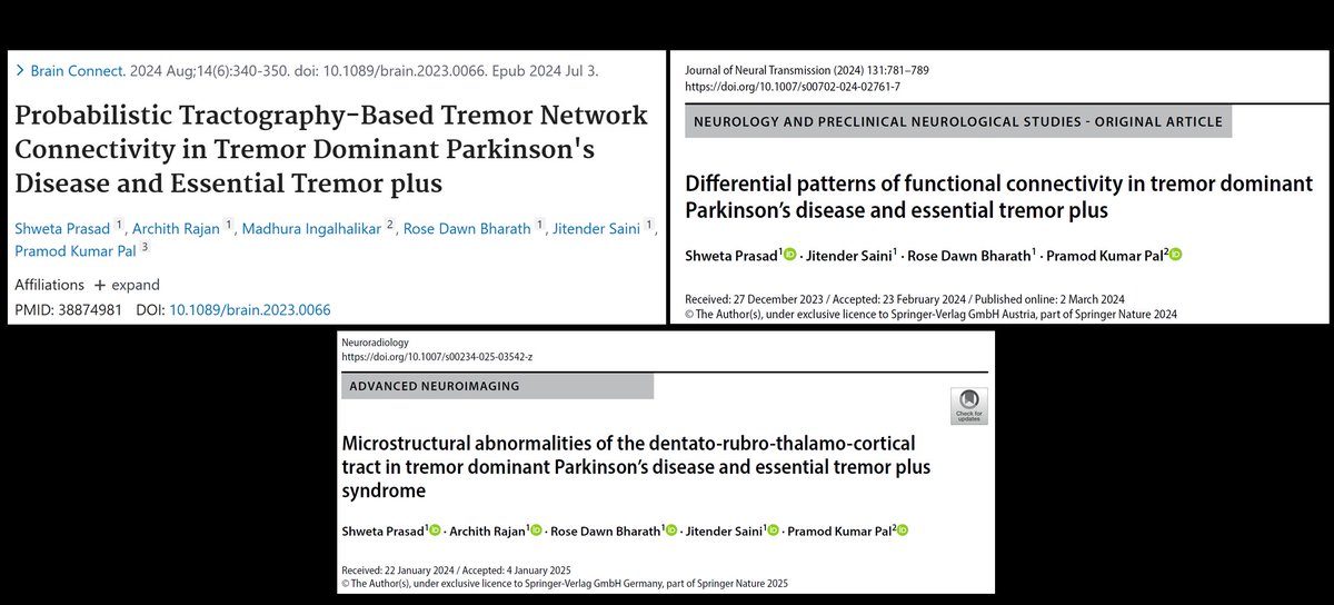
Glad to have been a part of the structural stuff.@ShwetaPrasad91 explores structural and functional sub-networks in Essential tremor and two sub-types of PD
This trifecta of publications is the outcome of several years of my obsession with tremor and neuroimaging. Rest assured, there's a whole lot more of where this came from.
The Spatial Organization of Ascending Auditory Pathway Microstructural Maturation From Infancy Through Adolescence Using a Novel Fiber Tracking Approach
https://t.co/SK0XD3ciXD
A major unmet need in #neurooncology is a sensitive, specific, reproducible imaging tool to differentiate #glioma progression (TP) from pseudoprogression (PsP).
https://t.co/ZeN149s8As
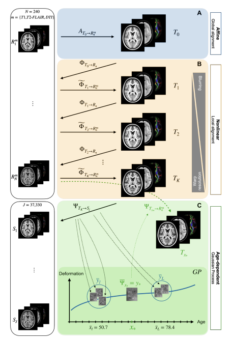
Internally-consistent and fully-unbiased multimodal MRI brain template construction from UK Biobank: Oxford-MM | Imaging Neuroscience | MIT Press https://t.co/oB3v1WeVQz