I've spun-out the framework used in our two-photon microscopy for cancer surgery into a set of packages called Dirigo: https://t.co/VCPw59MbkR
While it's a work in progress, I've been trying to write weekly-ish posts on design and features: Post #1 ...
@voigtvision@tripletimaging@jdwong_campos@Mackenzie_then This is very interesting but a potential concern is excess phototoxicity from intentional triplet state formation. Another thing is the observed power scaling of ~1.6 will be much worse than 2P in practice.
👀 Some of the proposed institute names are too general… National Institute on Body Systems Research? Some seem to lose the range of funded activities… NICHD to National Institute for Disability Related Research? 🤔 Others stay as is.
Full report: https://t.co/Li7F4VqYAQ
⚡️📣👇Tremendously excited to share our new @CellCellPress article, where we develop TriPath, a method for analyzing 3D pathology samples using weakly supervised AI. Article: https://t.co/7ydyLcPVww. TriPath enables 3D computational pathology via 3D multiple instance learning allowing AI models to capture intricate morphological details from pathology volumes.
Code: https://t.co/t9zeKWafsx
Blog post: https://t.co/pDzFMMG1qo
Tested on two different imaging modalities, and patient cohorts from two institutions. Our superstar @GreatAndrew90 put in a monumental effort of leading the study, in a fantastic collaboration with @jonliu123 at @UW .
Interesting aspects:
- Utilizing the whole tissue volume and leveraging 3D deep learning enable superior risk prediction performance compared to 2D deep learning baselines based on a few sampled tissue sections that emulate standard clinical practice. This indicates TriPath can harness additional information provided by 3D tissue morphology.
- The performance is also superior to clinical baselines from a reader study that involved six expert pathologists.
- The morphologically heterogeneous tissue volume could lead to opposing patient-level outcome predictions, dependent on which portion of the tissue volume is used. This concurs with current clinical literature warning that tissue sampling bias can lead to misdiagnosis.
Some limitations:
- While the 3D pathology cohort size is unprecedented, it is smaller than typical 2D pathology cohorts. Further large-scale studies will be required for validation. Nevertheless, we believe that this study will initiate a positive cycle, encouraging academic institutions and pharmaceutical companies to contribute large banks of human tissue blocks with paired clinical outcomes, thus speeding up advancements in 3D computational pathology.
Concluding insights: We believe that 3D pathology is just around the corner - It has the huge potential to not only augment/improve the current clinical practice centered around 2D examination of human tissue, but also help reveal novel biomarkers for prognosis and therapeutic response..
@harvardmed@harvard_data@MassGenBrigham@broadinstitute
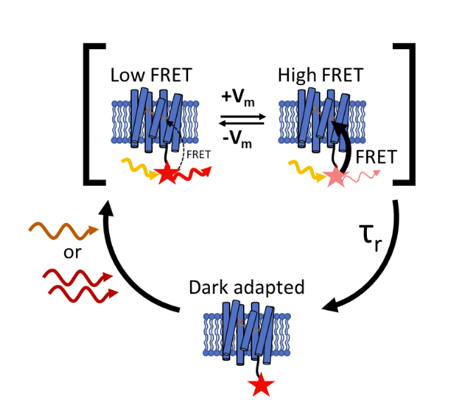
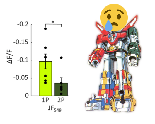
Their hypothesis: there is an intermediate state in the photocycle and that promotion to voltage-sensitive state requires excitation via longer wavelengths. 920/1040 nm 2P excitation used before is ineffective. They saw recovery to 1P-levels of voltage sensitivity at 1135 nm.
New preprint from @AdamEzraCohen's group shows that sensitivity may be recovered if longer wavelengths (beyond that of Ti:sapph, sadly) are used. https://t.co/BrabDY95lI
Voltron 1&2 are excellent voltage indicators, combining the sensitivity/targeting of genetically encoded microbial rhodopsin with the approaching-perfect photophysical performance of engineered organic dyes. However... (gif: https://t.co/3jnOF00wxb)
@voigtvision@Thorlabs@PodgorskiLab@brunopichler@Labrigger@ptrrupprecht Using them (an earlier eng prototype) successfully! As to whether they will work ok for you, depends on your photon rate and how efficient you need to be. It's definitely a pain to demag a 2 cm aperture onto a 3 mm detector. Look up Mike Giacomelli's papers, JBO and PLOS 1
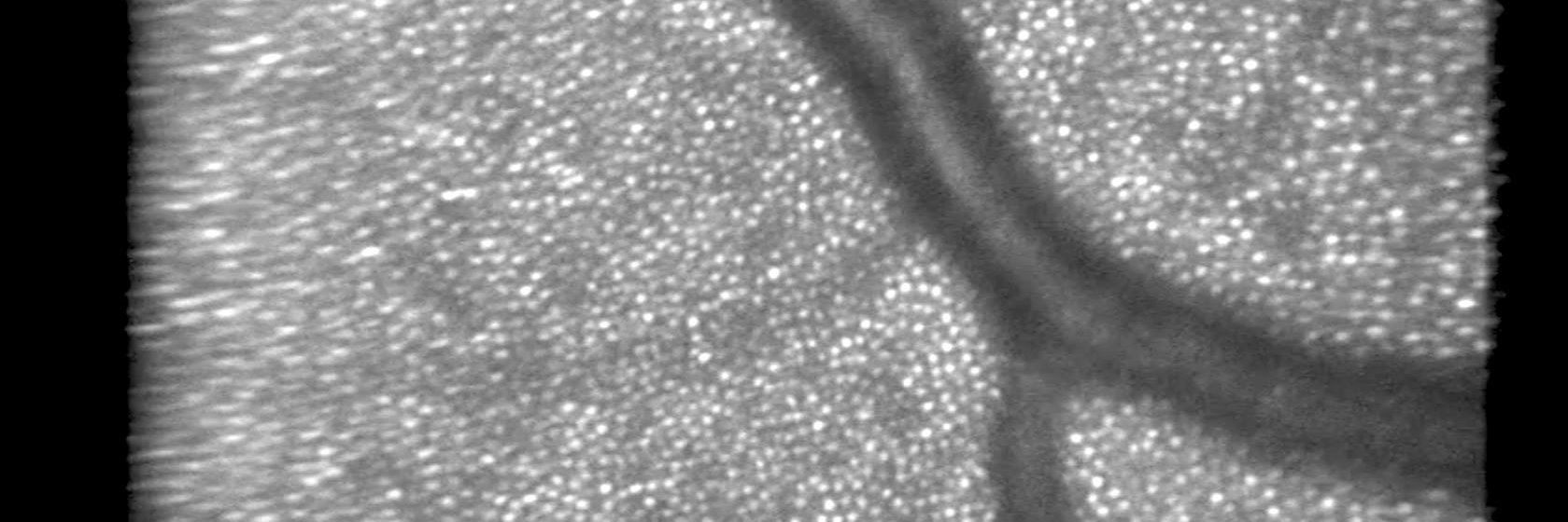
@Kevin_W_Bishop@NatureProtocols Amazing render! Is it max projection? When I've tried this on thick specimens max projection makes everything look way too hypercellular