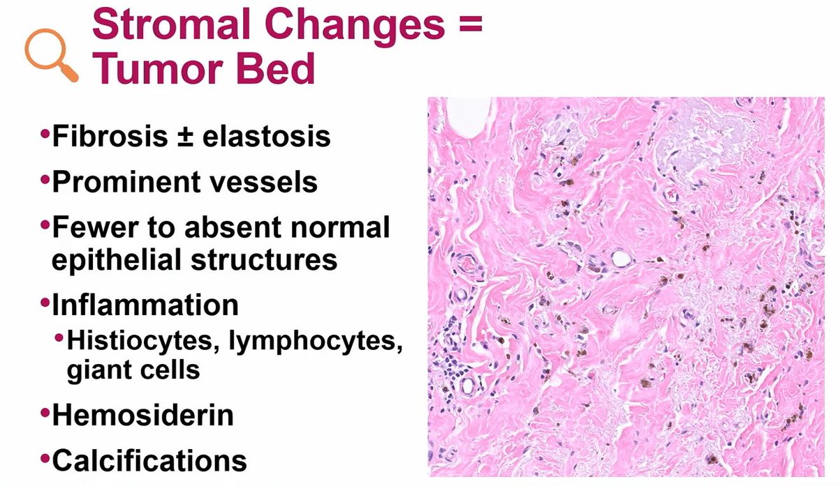
Workup of high-grade B-cell lymphoma/diffuse large B-cell lymphoma
- Establish cell of origin: Hans algorithm
- Assess for double-expressor lymphoma (DEL): MYC and BCL2 IHC
- Assess for double-hit lymphoma (DHL): FISH for MYC, BCL2, and BCL6
Dr. Sohani - 46th Annual Current Concepts in Surgical Pathology
If you submitted all the appendix and you don't see epithelium, just mucin in the lumen, sign it as:
-Dilated appendix with mucin
Comment: The differential includes LAMN or reactive processes
Dr. Umetsu #CAP23#everydayGI#GIpath#PathX#PathTwitter#pathology#pathologists
in #GUpath TURBT, areas suspicious for LVI are:
1) a great clue🧐for lamina propria invasion (stromal retraction)
2) infrequently represent actual LVI (CD31 #IHCpath shown here; not LVI)
Case of Papillary Endothelial Hyperplasia 🔬
Even blood vessels want in on the papillary party 🎉. Reminder that ‘papillary’ is about the pattern, not the cell type.
#PathX#PathTwitter#breastpath
#USCAP2025 w/ Dr Mete
Differentiated neuroendocrine neoplasms have lineage specificity- they replicate some of the developmental biomarkers based on their anatomic region or location. Contrast to NECs which ❌ respect lineage specificity (aberrant expression)
#PathWebinarPearls
📢 Half-Day Virtual Meeting
Topic: Syndromic Tumours in Gynaepath – Diagnoses Not to Be Missed by Pathologists!
🗓️ Join us for an engaging half-day online event.
💡 Gain key insights into recognising syndromic tumours in gynaecological pathology and ensure critical diagnoses aren’t overlooked.
🎟️ Early bird discounts are now available!
🎥 Can’t attend live? Enjoy anytime access for 12 weeks post-course.
To book 👉https://t.co/5WQSfSlKlE
#PathTwitter #Gynaepath #Gynpath
#USCAP2025 w/ Dr Voltaggio
Identifying 4 lines helps avoid missed dysplasia in upper GI & reduces equivocal cases → fewer interventions. Loss of lines = dysplasia.
4 Lines:
1.mucin cap
2.bottom of mucin cap
3.cytoplasm
4.nuclei
#PathWebinarPearls
Rules of well differentiated lipomatous neoplasms
1. Location (pic 2)
2. Do not look for lipoblasts (pic 2), look for atypical hypercromatic cells in fibrous septa (pic 3-you can see them at 10X, if you have to go to 40X to look for them then they are not real ones)
3. Cytogenetics are often helpful in appropriate classification (pic 4)
Dr. Fritchie- Comprehensive and Immersive Soft Tissue Pathology Course-May 2025 #pathology #BSTPath #PathX
Use of p53 immunohistochemistry can improve diagnostic agreement for differentiated vulvar intraepithelial neoplasia (dVIN): an international reproducibility study - Dasgupta - Histopathology - Wiley Online Library #gynaepath#p53#openaccess https://t.co/XHYAJq83tJ