Brasileiro perdido pelo mundo, buscando um sentido para a vida que vive dentro de mim.
Pai, Estomatologista, Patologista, cientista principiante e feliz.
The 2024 #NobelPrize laureates in chemistry Demis Hassabis and John Jumper successfully utilised artificial intelligence to predict the structure of almost all known proteins.
What groundbreaking discoveries will be recognised with a Nobel Prize this year? The announcements will begin in just a few days. Learn more: https://t.co/RCKc2r3asL
New in #HumPathol: Papillary Endothelial Hyperplasia in Late Post-Radiation or Late Post-Operative Breast Hematomas Mimicking Angiosarcoma. https://t.co/GGKuTactr7 #pathology#PathTwitter#PathX#breastpath
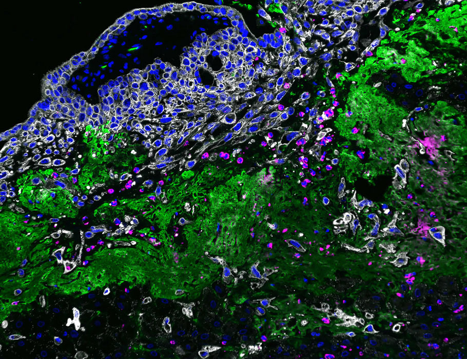
A new @SciImmunology study highlights a neutrophil subset that accumulates in the placenta and promote trophoblast invasion to support early human pregnancy. https://t.co/kgoQwV07JO
New in #HumPathol: Spatial and Interlesional Heterogeneity of FGFR2b Expression in Paired Primary and Metastatic Gastric and Gastroesophageal Junction Adenocarcinomas. https://t.co/UX0L9HXtQs #pathology#PathTwitter#PathX#GIpath
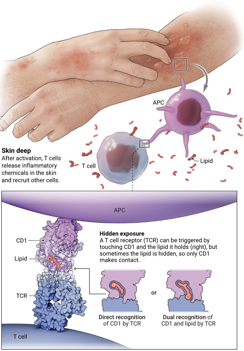
CD1 proteins on so-called antigen-presenting cells display lipids from pathogens to T cells to drive protective immune responses, but they also trigger skin inflammation in response to some lipids.
Learn more: https://t.co/c6l9t14uBR @NewsfromScience
Hoje dei uma aula sobre inteligência artificial e raciocínio clínico no programa teórico da Medicina Interna do HCFMUSP.
Falei sobre como essas ferramentas funcionam, possíveis usos na prática clínica e suas principais limitações.
https://t.co/WRLPNcwI6v
The number of female #BreastCancer survivors is projected to reach 5.3 million by 2035—an increase of one million women from 2025, marking the largest projected growth among the top 10 most prevalent cancers.
More stats at: https://t.co/53D4cKCXuS
@OncoAlert @docnikitawagle
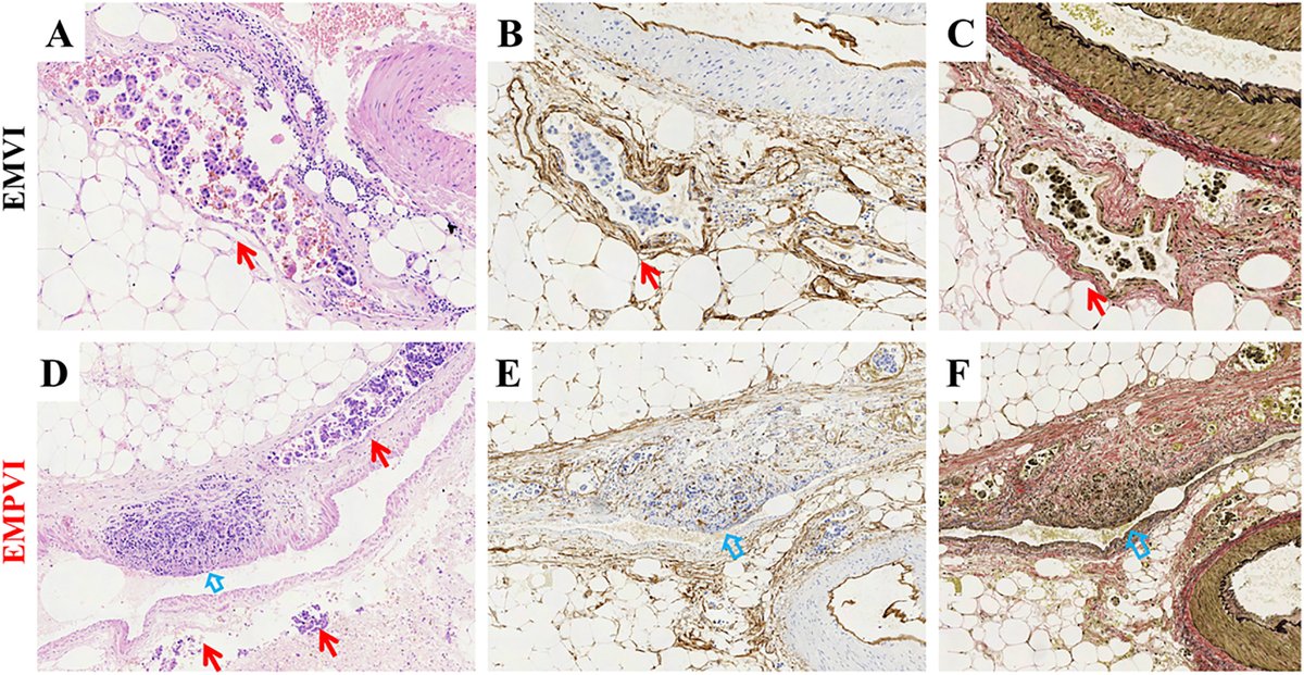
New in #HumPathol: Prognostic Value of the Extramural Perivenous Infiltration in Locally Advanced Gastric Cancer: A Retrospective Clinical Study. https://t.co/cbXHX9MDwP #pathology#PathTwitter#PathX#GIpath
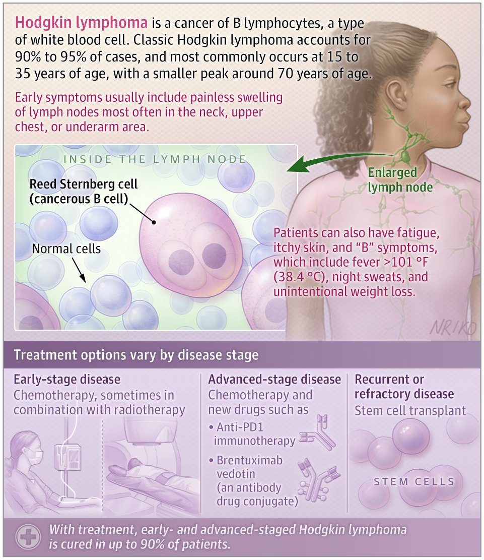
📝 #JAMAPatientPage: #HodgkinLymphoma is a cancer that arises from B lymphocytes, a type of white blood cell.
This JAMA Patient Page describes risk factors, common symptoms such as painless lymph node swelling and B symptoms, and how classic Hodgkin lymphoma is diagnosed and staged.
https://t.co/E8piXnXjdk
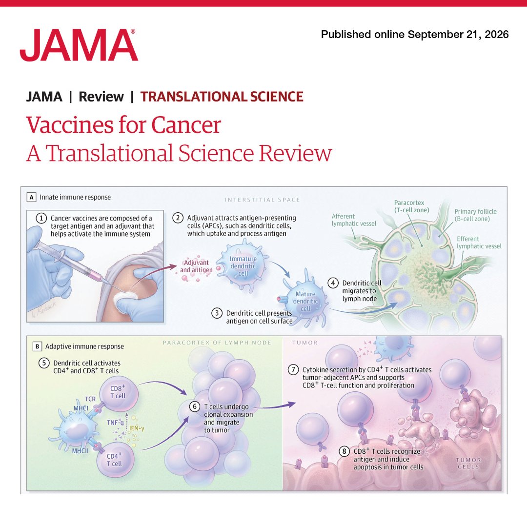
📝 #JAMAReview: #Cancer vaccines are being evaluated for treatment and prevention across multiple malignant neoplasms as advances in tumor immunology, antigen discovery, and vaccine delivery reshape the field.
This Review discusses how vaccines can stimulate adaptive immunity against neoantigens, nonmutated tumor proteins, or tumor-derived antigens, with current approved uses in melanoma, prostate cancer, and bladder cancer.
🔗 https://t.co/f4OJ6Qsyxx
Increased extracellular fluid viscosity drives M2-like macrophage polarization to promote breast cancer progression and immunotherapy resistance https://t.co/oqDMgguQJ5
🔬🤖 Pathology foundation models have transformed histology. Cytology has largely been left behind. CROWN may change that.
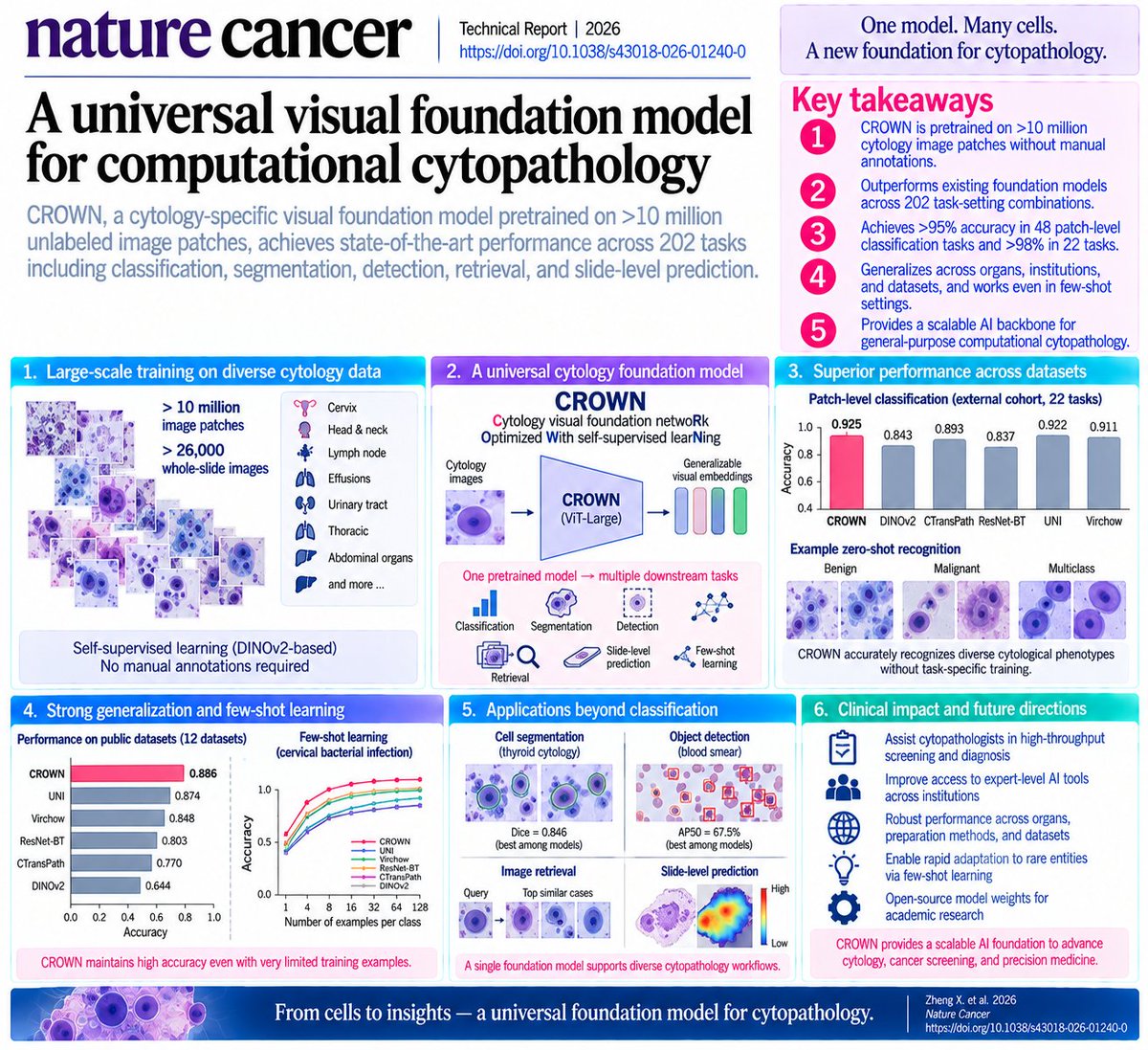
A new Nature Cancer Technical Report introduces CROWN (Cytology visual foundation netwoRk Optimized With self-supervised learNing)—a cytology-specific visual foundation model trained on >10 million image patches from >26,000 whole-slide images, without manual annotations during pretraining.
The scale of evaluation is particularly impressive:
202 task-setting combinations
spanning:
classification • image retrieval • segmentation • object detection • whole-slide prediction • few-shot learning
across multiple organs, institutions, preparation methods, and public datasets.
And the core finding is simple:
A foundation model trained specifically on cytology consistently outperforms general-purpose and histopathology foundation models on cytology tasks.
🧬 Why does cytology need its own foundation model?
Histopathology AI benefits from tissue architecture.
Cytology is fundamentally different.
Cells are obtained through minimally invasive sampling and appear as:
isolated cells + small clusters
without the spatial tissue architecture available in histological sections.
Yet cytology is enormously important clinically:
cervical screening
thyroid FNA
body-fluid cytology
urinary cytology
lymph-node cytology
bronchial specimens
and many others.
Existing AI approaches are typically trained for one narrow task.
CROWN instead asks:
Can one pretrained representation understand cytomorphology across organs and diseases?
🏗️ How CROWN was built
The model uses a:
ViT-Large
DINOv2 self-supervised student–teacher architecture
masked image modeling.
During training, 50–70% of image patches were randomly masked, forcing the network to infer missing morphological information from context.
Importantly:
No manual diagnostic labels were required for pretraining.
The training set covered cytology from:
cervix
head & neck
lymph node
effusions
urinary tract
thoracic sites
abdominal organs
and other clinical specimen types.
The overview on page 2, Figure 1 is worth seeing: it shows the >10-million-image training corpus feeding a single CROWN encoder that subsequently supports classification, retrieval, segmentation, detection and whole-slide tasks.
🎯 Zero-shot performance is already strong
The investigators first froze the pretrained representations and asked CROWN to recognize cytological classes without task-specific fine-tuning.
Across 34 internal patch-level tasks:
CROWN mean accuracy = 0.838
compared with:
DINOv2: 0.723
CTransPath: 0.778
ResNet-BT: 0.712
UNI: 0.835
Virchow: 0.820.
Then came the external cohort.
Across 22 classification tasks:
CROWN accuracy = 0.925
versus:
DINOv2 0.843
CTransPath 0.893
ResNet-BT 0.837
UNI 0.922
Virchow 0.911.
The feature-space maps on page 3 are visually striking: benign/malignant and multiclass cytological phenotypes form surprisingly clean clusters in the learned embedding space.
🔥 Give CROWN a little supervision—and performance rises further
With linear probing, only a simple classifier is trained while the CROWN encoder remains frozen.
Internal cohort:
Accuracy = 0.864
F1 = 0.854
Sensitivity = 0.753
AUC = 0.922
again outperforming the tested baselines.
In the external SCC cohort:
mean accuracy = 0.941.
Even more impressive:
15/17 binary tasks exceeded 95% accuracy
and
11/17 exceeded 98%.
This is not one cancer type or one specimen.
The benchmarks include cytology from:
pancreas
thyroid
cervix
urine
lymph nodes
bronchial specimens
pleural/pericardial/ascitic fluid
bile duct
breast
and others.
👀 What is the AI actually looking at?
The authors used Grad-CAM to visualize diagnostically important regions.
CROWN preferentially attended to classic cytopathological features including:
nuclear atypia
increased nuclear-to-cytoplasmic ratio
hyperchromasia
cytoplasmic vacuolation
cell clustering.
Two experienced cytopathologists independently reviewed the attention patterns, with substantial interobserver agreement.
The examples on page 5 are particularly compelling: heatmaps focus directly on abnormal cellular clusters in pancreatic, thyroid, lymph-node, pleural and other cytology specimens.
So the model is not simply generating accurate predictions.
Its attention overlaps recognizable human cytomorphological reasoning.
🌍 Perhaps more important: it generalizes beyond its training domain
CROWN was evaluated on 12 public cytology datasets.
Mean linear-probing accuracy:
0.886
versus:
DINOv2 0.644
CTransPath 0.770
ResNet-BT 0.803
UNI 0.874
Virchow 0.848.
AUC reached:
0.960.
Then the investigators deliberately tested an out-of-distribution domain absent from pretraining:
🩸 peripheral blood smears.
CROWN still achieved:
accuracy 0.848
sensitivity 0.908
AUC 0.906.
That is exactly the kind of generalization a foundation model is supposed to provide.
🔎 CROWN can also function as a cytology search engine
This is an underappreciated application.
A pathologist can theoretically provide a query image and retrieve morphologically similar examples.
CROWN was tested for:
anatomical-site retrieval
and
fine-grained diagnostic retrieval.
In the internal cohort, 1,887 query images were searched against 17,154 candidate images.
In the external cohort:
1,378 queries
against
12,527 images.
CROWN achieved the highest retrieval performance at both anatomical and subtype levels.
The examples on page 6 make the potential obvious:
query cell image
→ CROWN embedding
→ nearest morphologic neighbors
→ Top-1 / Top-3 / Top-5 comparable cases
This could eventually support:
case comparison
education
rare-disease retrieval
and
diagnostic decision support.
🧫 It isn't just classification
The same backbone supports cell segmentation.
For thyroid cytology:
DICE = 0.846
and cervical cytology:
DICE = 0.719
both the highest among the tested encoders.
For blood-cell object detection, CROWN achieved:
AP50 = 67.5%
compared with:
DINOv2 44.1%
CTransPath 33.1%
ResNet-BT 53.3%
UNI 64.8%
Virchow 44.9%.
The segmentation/detection comparisons on page 29 reinforce an important point:
one pretrained backbone → many different cytopathology tasks.
🔬 And it works at the whole-slide level
The authors then tested 12 weakly supervised slide-level tasks involving:
2,817 unique slides
and
5,190 slide-task instances.
CROWN achieved:
mean accuracy = 0.794
AUC = 0.875
sensitivity = 0.741.
For binary tasks alone:
mean accuracy = 0.867.
One particularly interesting example:
primary origin of lymph-node metastases
—a five-class problem—
achieved:
90% accuracy.
The slide-level Grad-CAM examples on page 8 show CROWN identifying diagnostically informative microscopic regions within huge cytology slides rather than treating the entire WSI uniformly.
🤯 Few-shot learning may be the most clinically useful feature
Rare cytological entities create a fundamental AI problem:
there simply aren't thousands of labeled examples.
So the investigators tested CROWN with only:
1
4
8
16
32
64
128
or
256
slides per class.
Across few-shot evaluations:
CROWN mean accuracy = 0.620
and it outperformed every comparator.
For cervical bacterial infection detection:
only 8 examples/class → accuracy 93.5%.
None of the comparator models exceeded 80% even with 128 examples.
The learning curves on page 10 make this especially striking: CROWN separates from the competing foundation models as the number of available examples increases.
This may be crucial for:
rare cancers
rare cytological subtypes
small institutional datasets
where conventional supervised AI development is impractical.
🧠 Why does a cytology-specific foundation model matter?
Histology foundation models such as UNI and Virchow were trained on enormous pathology datasets.
But histology asks the model to understand:
cells + glands + stroma + tissue architecture.
Cytology asks something different:
nuclear morphology
chromatin texture
N:C ratio
cell clustering
cytoplasmic morphology
isolated-cell phenotypes.
CROWN's results suggest that:
domain-specific pretraining matters.
A model trained explicitly on cytology can outperform even much larger histopathology foundation models on cytological problems.
The authors themselves note that the largest gains occur in high-level semantic tasks such as:
classification
detection
retrieval,
whereas gains in lower-level segmentation are smaller.
That distinction is informative about what the foundation representation is actually learning.
🚀 The future workflow could look very different
Imagine:
FNA / brushing / urine / effusion / cervical smear
↓
digital image
↓
CROWN
↙︎ ↓ ↘︎
classification
abnormal-cell detection
segmentation
similar-case retrieval
rare-disease few-shot classification
↓
cytopathologist review
↓
AI-assisted diagnosis
Importantly, the authors even note that patch-level images were captured directly through routine microscopes—not exclusively through expensive whole-slide scanners.
That could eventually make this architecture relevant to laboratories without comprehensive digital-pathology infrastructure.
⚠️ But CROWN is not yet a universal clinical cytopathologist
There are important limitations.
The large training corpus came from a limited number of institutions and geographic regions, so true global generalizability remains uncertain.
CROWN is also currently:
VISION ONLY.
It does not yet integrate:
clinical history
cytology reports
immunocytochemistry
molecular testing
genomics
which are often essential to real cytopathology diagnosis.
And although the benchmarking is extensive, the study is retrospective and computational. The authors report that no formal statistical hypothesis testing was performed for comparisons between models.
Prospective workflow studies are therefore the real next step.
🎯 The larger message
The major contribution may not be another model that achieves slightly better accuracy.
It is the demonstration that:
>10 million unlabeled cytology images
↓
self-supervised learning
↓
ONE FOUNDATION MODEL
↓
zero-shot diagnosis
supervised adaptation
image retrieval
segmentation
cell detection
whole-slide analysis
few-shot rare-disease learning
can work across remarkably heterogeneous cytological specimens.
That changes the engineering paradigm.
Instead of building:
one AI model → one organ → one diagnostic task
we may increasingly build:
ONE CYTOLOGY FOUNDATION MODEL
↓
many organs × many diseases × many tasks.
And because CROWN's code and model weights are available for academic research, this is not merely a conceptual demonstration—it is a foundation other groups can directly build upon.
For anyone working on AI + minimally invasive cancer diagnostics, this is a model worth watching.
📄 Zheng X, Zheng K, Wang J, et al.
A universal visual foundation model for computational cytopathology.
Nature Cancer. 2026.
DOI: 10.1038/s43018-026-01240-0
#ArtificialIntelligence #Cytopathology #DigitalPathology #FoundationModel #CROWN #CancerDiagnostics #ComputationalPathology #DeepLearning #DINOv2 #PathologyAI #PrecisionOncology #CancerScreening #MedicalAI #NatureCancer
#ScienceSaturday What if #cancer starts weakening T cells before they even reach the tumor?
A new study published in @Nature suggests that an important part of cancer’s immune suppression may happen outside the #tumor itself, in nearby lymph nodes where cancer-fighting T cells are activated and prepared to respond.
🔬 Researchers studied tumor-draining #lymph nodes from patients with melanoma and identified a population of immune cells, mainly macrophages, producing high levels of an enzyme called PLA2G2D. These cells were found close to activated CD8+ T cells and were associated with poorer outcomes.
In laboratory and mouse studies, PLA2G2D acted like an immune brake, limiting the early expansion of tumor-specific T cells. When researchers blocked it, more cancer-fighting T cells expanded in the lymph nodes, entered the bloodstream and ultimately reached the tumor.
Even more interesting, blocking PLA2G2D alongside PD-1 checkpoint inhibition produced additive or synergistic antitumor effects in preclinical models, suggesting the two pathways may suppress #Tcells in different ways.
💡 Why does this matter? We often focus on overcoming immune suppression inside the tumor. This study suggests there may also be important immune “checkpoints” in the lymph nodes where antitumor responses begin.
There is still more work to do before this approach can be tested as a treatment in patients, but understanding these early barriers could reveal new ways to help more tumor-specific T cells make it into the fight. @AvanKrimpen_@R_Stadhouders@bart_lambrecht@ErasmusMC@tmu2010@UTokyo_News_en@NatureComms@NatureMedicine
Read the study: https://t.co/qRABB2nQpo
Introducing Jev Reviewer, a tool designed for systematic reviewers to quickly find and extract the information from research articles.
App: https://t.co/ScFHy5dYFR
GitHub: https://t.co/gaTlohbGc1