The nuclei of the cells in the heart of a zebrafish embryo photographed through a microscope. The ventricle is on the left and the atrium is on right. #CellBiology
🎄✨ Merry Christmas! 🎅🏻 Wishing you all a day filled with love, laughter, and the warmth of cherished moments. May your hearts be as full as your plates and your spirits as bright as the twinkling lights.
Apply for an exciting opportunity to visit the Kawakami Lab through the National Institute of Genetics Travel Grants. The deadline for the application is December 1, 2023 (Japanese time). Learn more about this grant and apply here: https://t.co/4eqkwIk2FA
A time lapse of a cell going through cell division hand drawn by Walther Flemming while he was looking in his microscope in the 1800's. He chose to highlight the dynamics of chromosomes.
Our mesoscopic oblique plane microscope is online:
https://t.co/ij8xd8iibn
It enables rapid volumetric imaging of a whole zebrafish embryo with ~2.3 micron lateral resolution.
This is enabled by merging a couple of optical innovations and tricks. 1/N
Where do #TissueTregs come from? The previous standard model is the "seeding and specialisation" model, where #Tregs enter from tissues, turn on a dedicated transcriptional program per #tissue, and dwell indefinitely in that tissue as specialised cells.
1/3
FDA Approves Mirikizumab, a Promising Induction and Maintenance Therapy for Ulcerative Colitis. Offers new option with improved quality of life for patients with moderate-to-severely active disease https://t.co/IJoYYifqCa
Hi Twitter! I have a PhD!
I have many, many people to thank for getting me to this point, but most especially @JudyCho7 and @Felix_Chuang240 . I am so fortunate to have your mentorship and support.
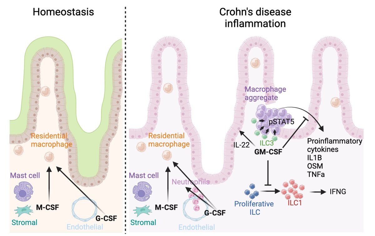
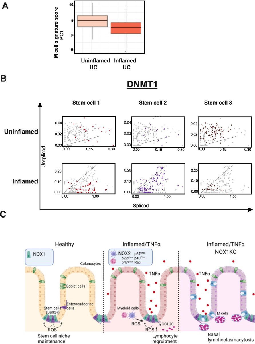
Our new paper is out @Gut_BMJ today. ROS is crucial for maintaining epithelial stem cells. Lossing ROS results in #M cell expansion and basal #lymphoplasmacytosis.