You can find our technologies in China! Martin is there with our new machine for Freeze substitution❄️ without liquid N2! (please see the second and third photos). Can't wait to deliver it to you! #EM#CLEM#electronmicroscope@AcadSinica
Max’s review, “Stress granules as causes and consequences of translation suppression” is online!
#stress#RNA#translation @maxbaymiller
Access on our website: https://t.co/LqdBs3Dja2
Our work describing Chromatin Expansion Microscopy is now out in @ScienceMagazine https://t.co/ysL8Q1xmqA Check out how we were able to visualize transcription, chromatin, and TFs with molecular resolution in developing zebrafish embryos to understand gene regulation!
Three #PhD positions open
@ucddublin to demonstrate benefits of #SXT in Correlative Imaging #CLEXM for disease research and drug delivery. The project is funded by #MSCA Doctoral Networks. Apply via https://t.co/hcaiBxzwG9 #PhDposition 🙏Please retweet
☕️NEW: @FrydmanLab@EmSontagMU@FaboPolanco & co show that nuclear & juxtanuclear misfolded proteins converge to opposite nuclear envelope sides, proximal to nuclear–vacuolar junctions for ESCRT-dependent egress to the vacuole.
https://t.co/DdNfaP1LNf https://t.co/EGfd9dKtuP
Out today at Nature Cell Biology our exciting story defining the coordination between cytoplasmic and nuclear spatial protein quality control: https://t.co/m5SgoEtZk9. Congratulations to @EmSontagMU and @FaboPolanco !!!
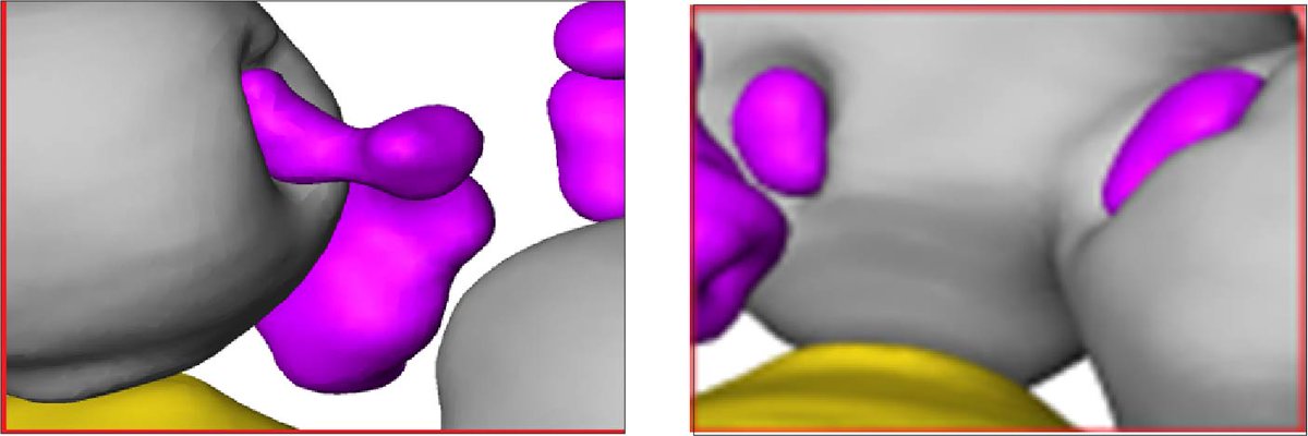
It could! We see cytoplasmic misfolded protein compartments interacting with vacuoles and deforming the vacuolar membrane, almost like they are being pulled in to the vacuole. #proteinclearance#autophagy#vacuole 5/12
IT'S FINALLY HERE! A story years in the making about how cells handle misfolded proteins in the nucleus and cytoplasm 1/12
#proteostasis#proteinfolding https://t.co/2F6cRe7Yti
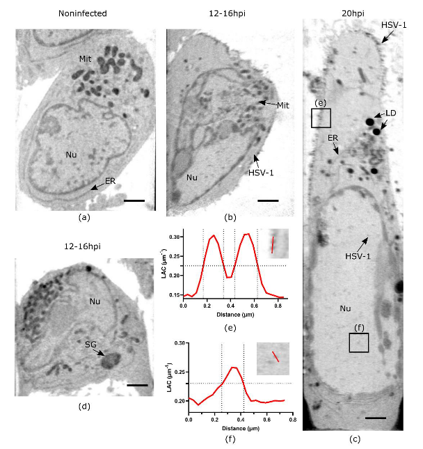
Soft X-ray #Tomography Reveals #HSV-1-Induced Remodeling of Human B Cells
Chen et al. demonstrate the power of #SXT imaging for scouting #virus-induced changes in infected cells and understanding the orchestration of virus-host remodeling quantitatively
https://t.co/8q2Y2vNg6U
Excited to share our new preprint is out! We're exploring the utility of X-ray tomography for virus research 🦠🧪. Collab between @AtomicVirology, @B24Light and the Colin Crump Lab.
A tweetorial (1/12)
https://t.co/XCachAh8dh
Excited to see our SARS-CoV-2 syncytia paper finally out in eLife. Congrats to @DavidWSanders2@LeventalLab@PlossLab and all our amazing collaborators: https://t.co/HFS53ze3Et
An exciting collection on Phase Separation in Biology from our colleagues at @NatRevMCB . And it includes one of our papers: "The Structural Basis of Rubisco Phase Separation in the Pyrenoid" from @JonikasLab
https://t.co/q1VCkKryYn
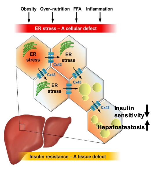
New paper on a groundbreaking study of stress response in liver cells is out in @Cell_Metabolism Congratulations to Gökhan S. Hotamışlıgil, Amir Tirosh, Gürol Tunçman, Ediz Calay, and others for their wonderful work. @ghotamis@MET_HSPH@HarvardChanSPH
https://t.co/IfCyKb5gsp
After years of hard work, Güneş @gparlakgul & Ana @apsarruda have successfully produced spectacular resolution of the inner architecture of liver cells, compared lean/obese liver tissue, & demonstrated the impact of architecture on metabolic function.https://t.co/MuaYlCbmxF
Excited to share the Preprint in collaboration with @MitoPsychoBio «3D Neuronal Mitochondrial Morphology in Axons, Dendrites, and Somata of the Aging Mouse Hippocampus» https://t.co/J4jQ9QaDCZ
@MitoResearch@mitomusclelab@EMRSncl
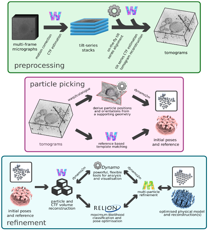
Optimal cryo-ET and subtomogram averaging workflows can be a pain - we put together a little toolkit which simplifies working between Warp-RELION-M and Dynamo, providing automation where possible and more flexibility if needed! 1/n https://t.co/1lFQ4XGlvc