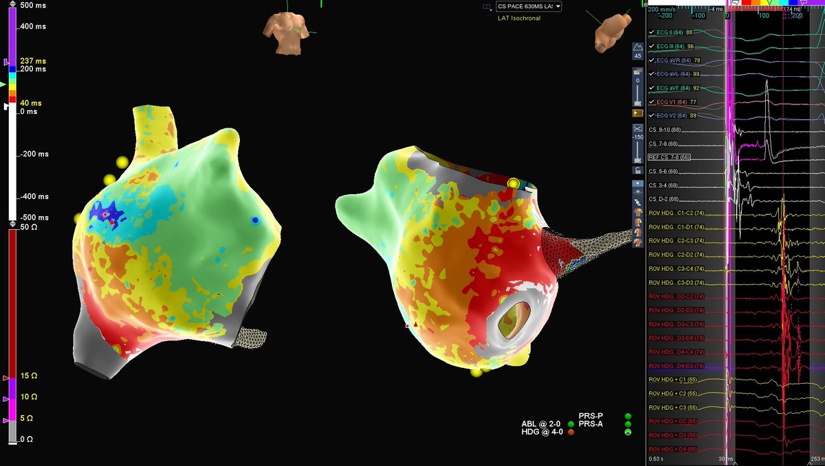
Case after case - Wavefront Direction is Key: Pacing perpendicular to WADLs is best for identifying fixed lines of block that perfectly match the VT isthmus lateral boundaries. @BIDMC_VT with star fellow Gabriel Odozynski, maps with @sarah_chomos#ablateVT
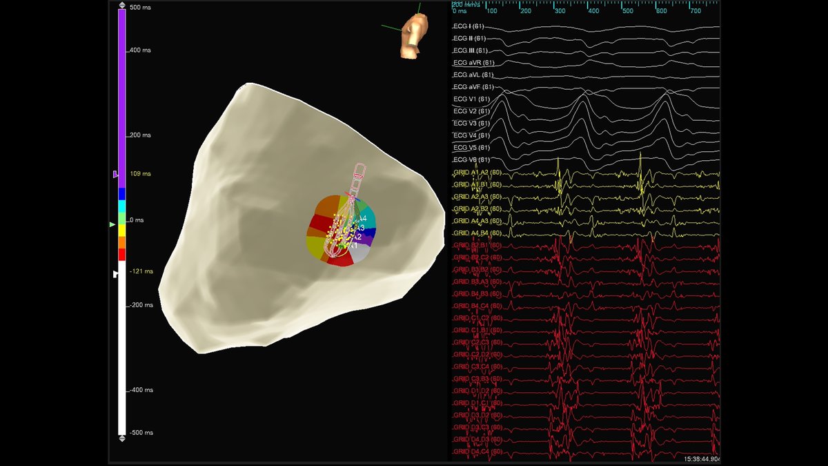
With multi-wavefront mapping in VT ablation we are always looking for new ways to visualize core concepts. Emphasis mapping on the activation window with ILAM highlights the narrowest isochrones to visualize WADLs which co-localize with the 3D boundaries of the VT. (Thread 1/6).
Remarkable agreement between ILAM deceleration zone, CT wall thickness channel, and peak frequency emphasis on the apical scar. Case by @davilandre and Dr. Bahij Kreidieh. Ensite mapping by @DavisSneider with @ADAS3D integration. #AblateVT
#OTNF annotation used to automatically display fascicular conduction AND slow conduction (DZs) in ICM w septal disease on a single map. No dynamic windowing, no manipulation. Just smarter annotation, cleaner maps and more informed decision making. @JRWinterfield@MUSC_EP
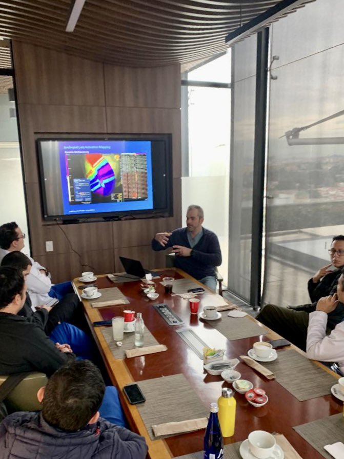
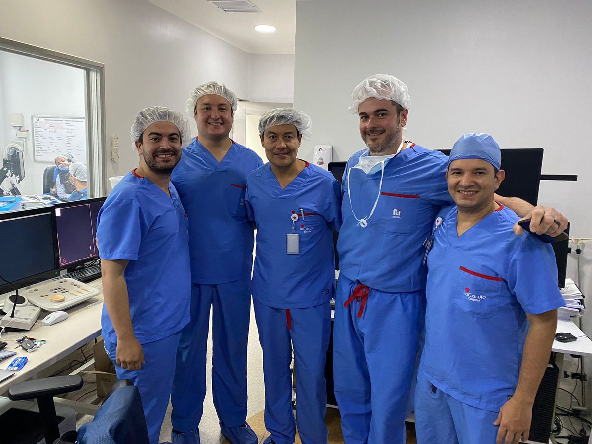
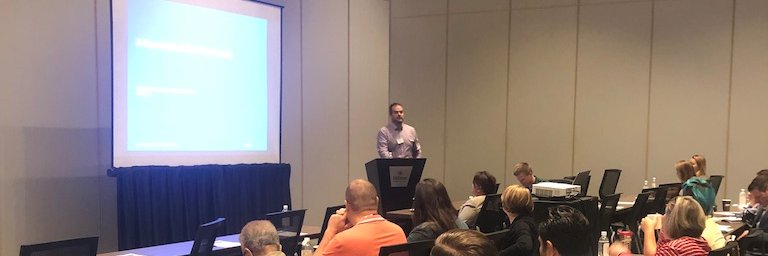
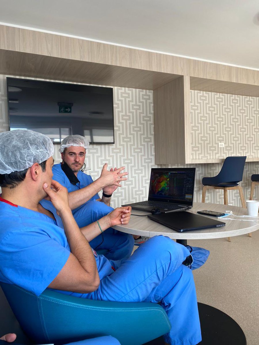
What a wonderful week of learning and sharing information with the incredibly talented and thoughtful team @fcardioinfantil pushing the field of EP to serve their patients! It was an honor to share the strengths that #EnsiteX will bring to this world-class center of excellence.
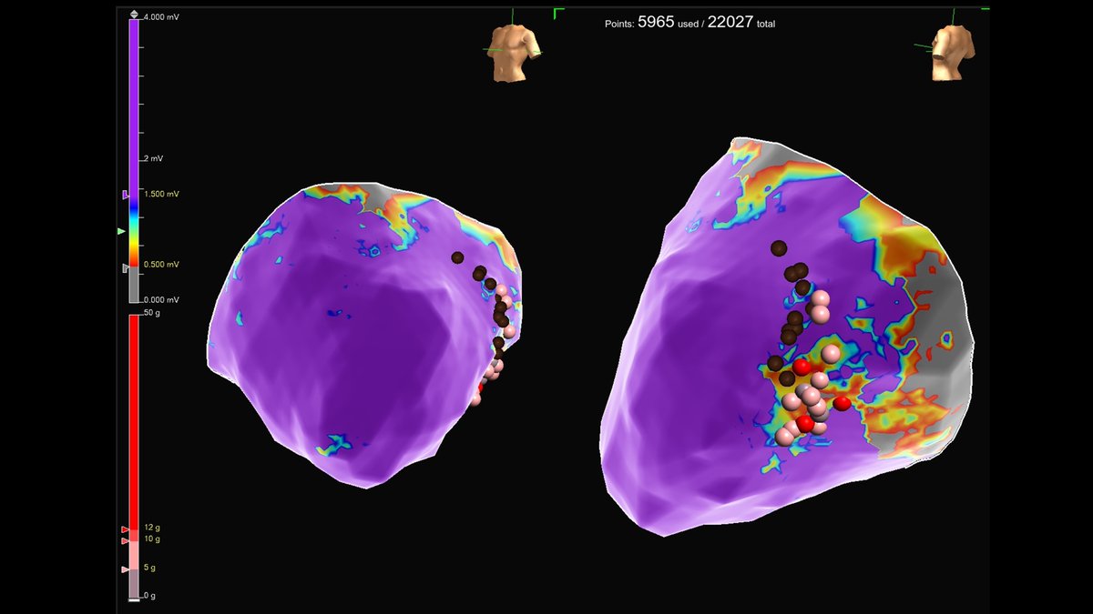
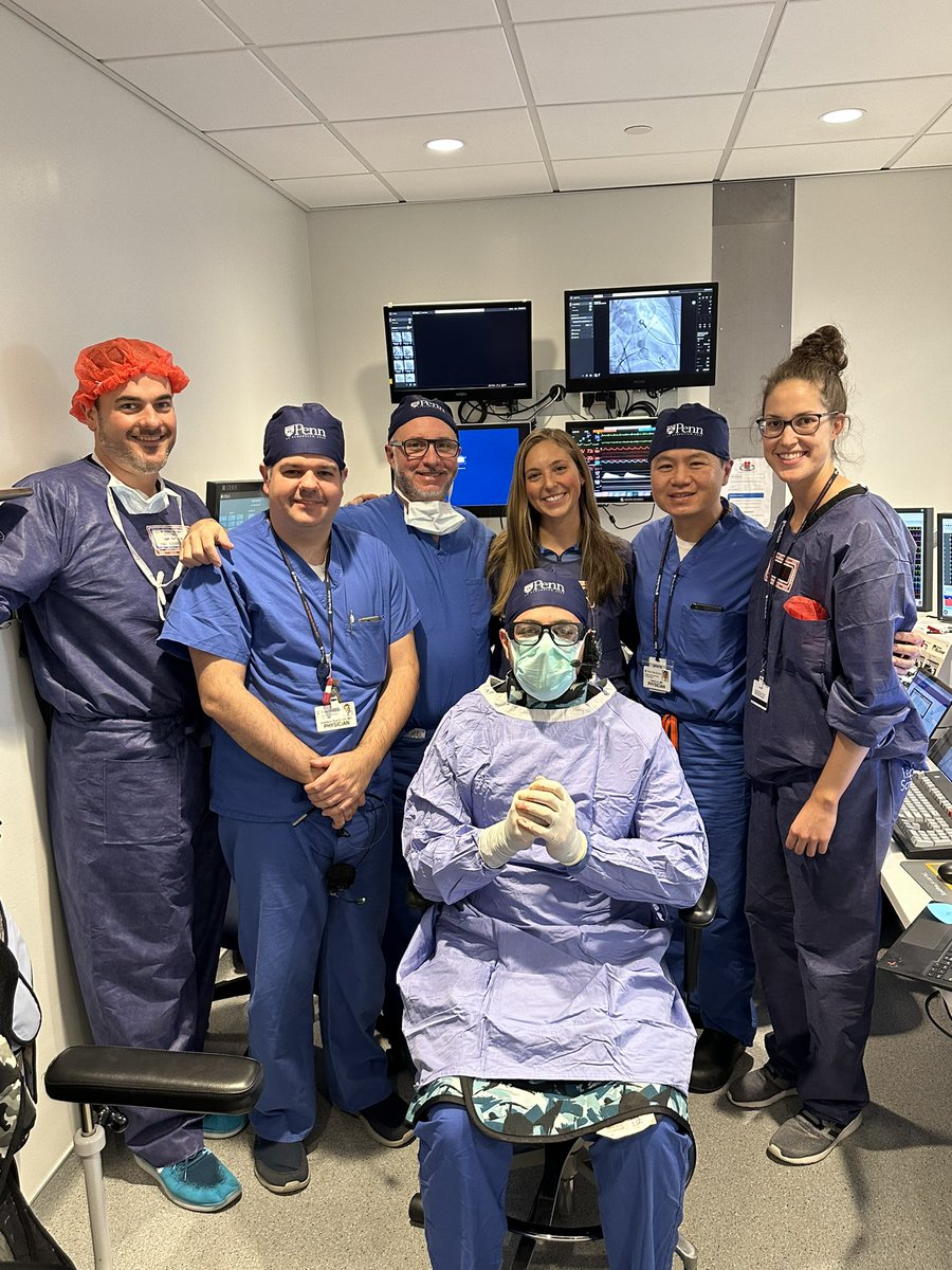
Patient (FLNC mutation) with ICD shocks despite AAD. PES with HD grid at site of epi low voltage , most of the VT circuit captured. Ablation epicardial unfortunately limited by phrenic and coronaries. Consolidated on endocardial aspect.
#EPeeps#ablateVT@PennCardiology
Want to highlight efforts of former fellow, and exceptional EP @Hkardioep who spearheaded this effort, and the mapping talents of @reedbonvo_EP and @lib_rosinski
#EPeeps A fairy tale of ILAM, coauthored with @cm_beach and illustrated by @TimStevens09 🧵
Chapter 1: Once upon a time, there was a humble deceleration zone. Although it was small, it seemed to have a lot of potential.