In 2016, two landmark studies showed that synchronous endometrioid endometrial and ovarian carcinomas, often classified as independent primaries, are actually clonal in the majority of cases
Evidence suggests these tumors are of endometrial origin with secondary ovarian involvement
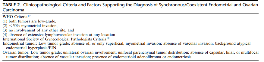
Despite their clonal relationship, prognosis can be excellent when “low-risk” features are present (see pic below)
FIGO 2023 endometrial carcinoma staging now includes substage IA3 for low-risk coexistent endometrial and ovarian carcinoma (CEOC)
If a CEOC does not meet low-risk criteria, FIGO recommends staging it as endometrial carcinoma with spread to the ovary
Rarely, patients have truly unrelated synchronous tumors (e.g., endometrioid endometrial carcinoma + incidental ovarian mucinous borderline tumor/carcinoma). These are staged and managed as independent primaries and are not included in the CEOC category
You can check different CEOC presentations and classification: Huvila et al. Coexistent Endometrial and Ovarian Carcinoma: Terminology, Clinicopathological Features, and Clinical Significance. DOI: 10.1097/PGP.0000000000001177
#GynPath #Pathology #PathX
Breast-Lobular vs ductal
- 3x fewer high-risk Oncotype DX scores but 5 year disease free survival similar to IDC NST
- Lobular less likely to have pathologic complete response (pCR) after neoadjuvant chemotherapy
- More likely to have HER2 mutations in the metastatic setting and benefit from targeted therapy may be greater in patients with ILC
Dr. Calhoun #BreastPath #pathology #USCAP #PathX
Interdigitating reticulum cell sarcoma can be considered a malignant variant of Rosai-Dorfman disease! S100 protein was + (not shown).
Ravindran A, et al . Mod Pathol. 2023 Oct;36(10):100268. PMID: 37406859.
Epithelioid hemangioma / Angiolymphoid hyperplasia with eosinophilia (with emphasis on diagnostic pitfalls)
Benign vasoproliferative lesion, rare, typically located in the head and neck, which can mimic malignant vascular tumors and Kimura disease – so it deserves a fixed place on the radar of dermpath and surgeons.
Definition 🧾
Benign vasoproliferative lesion composed of proliferating blood vessels lined by epithelioid/histiocytoid endothelium, associated with a lymphocytic infiltrate rich in eosinophils and often lymphoid follicles.
In contemporary literature, “angiolymphoid hyperplasia with eosinophilia” and “epithelioid hemangioma” are used as synonyms for the same process.
Epidemiology 📊
Rare, mainly affects young and middle-aged adults (mean 30–40 years), with no clear sex predominance in large series.
About half of patients present with a single lesion and half with multiple lesions; it is not considered an endemic disease, although more frequently reported in the Northern Hemisphere.
Sites 📍
Predominantly involves skin and subcutis of the head and neck, especially the periauricular region, face, and scalp.
Less common sites include trunk, extremities, penis, female genitalia, oral mucosa, and, rarely, deep locations (medium-sized arteries, bone).
Pathogenesis 🧬
Multifactorial pathogenesis, with three main lines of interpretation: reactive lesion after trauma/arteriovenous shunt, benign vascular neoplasm, and T‑cell monoclonal lymphoproliferative process with secondary angiogenesis.
Reported features include acquired arteriovenous shunts, monoclonal T‑cell receptor rearrangement in a subset of cases, and isolated somatic mutations in TEK (TIE2), with hormonal factors, atopy, and viral infections proposed as additional cofactors.
Clinical Features 🩺
Firm dome-shaped papules or nodules, pink‑erythematous to violaceous, ranging from a few millimeters to several centimeters; when clustered, they may form nodular plaques with a “grape-like” appearance.
Common symptoms include pruritus, easy bleeding, and pain or tenderness; some patients are asymptomatic, and a palpable pulsation can be detected in lesions with high-flow components.
Laboratory Diagnosis 🧪
Peripheral eosinophilia and elevated IgE occur in a minority of patients (around 20%), much less frequently than in Kimura disease.
Regional lymphadenopathy, when present, is usually reactive; the clinical course is chronic and indolent, with no spontaneous regression in most cases.
Histopathology 🔬
🖋️Architecture: dermal-based lesion often extending into the subcutis, ranging from well-circumscribed nodules to more diffuse proliferation within fibromyxoid stroma.
Endothelial cells: plump cells with eosinophilic, sometimes vacuolated cytoplasm, vesicular nuclei, epithelioid/histiocytoid or “hobnail” morphology protruding into the lumen, without marked cytologic atypia.
🖋️Inflammatory infiltrate: dense lymphocytic and plasmacytic infiltrate with lymphoid follicles and numerous eosinophils, which may account for more than 50% of the infiltrate in classic lesions.
🖋️Stroma and vessels: fibromyxoid stroma with hemorrhage, hemosiderin deposition, organized thrombi, and, in many subcutaneous lesions, altered arteries and acquired arteriovenous shunts.
Immunohistochemistry 🧫
🟢Positive: strong and diffuse CD31 expression, CD34 usually positive, von Willebrand factor variably positive, and intense nuclear FOSB expression in many cases.
🔴Negative/auxiliary: HHV8 negative (helpful to exclude Kaposi sarcoma); histiocytic markers such as CD68 are negative in endothelial cells despite their “histiocytoid” morphology.
Other: monoclonal T‑cell receptor rearrangement can be detected in a subset of cases, supporting an associated T‑cell lymphoproliferative component.
Differential Diagnosis 🎯
Kimura disease – deep subcutaneous masses, frequent lymphadenopathy and much more pronounced peripheral eosinophilia/IgE elevation; histology dominated by exuberant lymphoid follicles with eosinophilic microabscesses and less prominent epithelioid vascular proliferation.
Cutaneous angiosarcoma (including epithelioid variant) – elderly patients, infiltrative and aggressive lesions with anastomosing vascular channels, marked nuclear atypia, atypical mitoses, and necrosis, which are absent in epithelioid hemangioma.
Epithelioid hemangioendothelioma – low‑grade malignant vascular tumor with cords and nests of vacuolated epithelioid cells in a myxohyaline stroma, usually lacking a prominent lymphocytic/eosinophilic infiltrate and often harboring WWTR1::CAMTA1 rearrangement.
Kaposi sarcoma – HHV8‑positive, with slit‑like vascular spaces, fascicles of spindle cells, and hyaline globules; epithelioid hemangioma is HHV8‑negative and shows epithelioid endothelium with dense lymphocytic/eosinophilic inflammation.
Pyogenic granuloma – common rapidly growing, polypoid lesion with lobular capillary architecture, without prominent epithelioid endothelium or marked tissue eosinophilia.
Prognosis 📈
Benign lesion with no documented metastatic potential, but with a chronic course and high local recurrence rate, especially after incomplete treatment.
The main morbidity is cosmetic and psychosocial; extensive, multifocal, and repeatedly recurrent disease may require long-term follow‑up and multimodal management.
Treatment 💊
Surgical excision is the most commonly used modality and shows the lowest recurrence rate compared with other options, although recurrences still occur.
Vascular lasers (pulsed dye, CO₂ and others) are important alternatives in patients with multiple lesions or involvement of cosmetically sensitive areas; topical/systemic corticosteroids alone have high failure rates.
Other reported therapies include topical immunomodulators, beta‑blockers, and various systemic agents, mostly described in small series or case reports, with variable responses.
Take-Home Messages @Notas_Patologia 📌
🧩Epithelioid hemangioma/angiolymphoid hyperplasia with eosinophilia is a benign but locally recurrent vasoproliferative lesion, classically affecting the head and neck.
🧩The combination of epithelioid endothelium and dense lymphocytic infiltrate rich in eosinophils is the central histologic clue.
🧩Distinguishing it from Kimura disease and malignant vascular tumors (angiosarcoma, epithelioid hemangioendothelioma) is crucial to avoid overtreatment.
🧩Nuclear FOSB expression and HHV8 negativity are useful immunohistochemical features, especially when Kaposi sarcoma and other vascular tumors are in the differential.
🧩Complete excision (sometimes with Mohs or laser) is the mainstay of therapy, but patients should be counseled regarding the risk of local recurrence.
Selected References 📚
WHO Classification of Skin Tumours, latest edition.
Olsen TG, Helwig EB. Angiolymphoid hyperplasia with eosinophilia. J Am Acad Dermatol.
Adler BL et al. Epidemiology and management of epithelioid hemangioma: systematic review of 908 cases.
PathologyOutlines – Epithelioid hemangioma / Angiolymphoid hyperplasia with eosinophilia.
⚠️Disclaimer⚠️
“This text is an educational summary for healthcare professionals and students. It does not replace full pathology reports, local guidelines, or individualized clinical decision-making.”
Hashtags
#MedicalEducation #NotasDePatologia #pathology #Dermatopathology #VascularTumors
#GUPath#Surgpath#Pathtwitter#PathX#Pathresidents: Let’s take a look at two biopsy cores (from the same patient) and decide what to call them.
Take a look at both cases (“A” in this post, “B” in reply). Cast your vote. Then, read on for some teaching points geared towards trainees and general fans of #GUPath.
What about WHO5?
The data seems to suggest that MYC + BCL6 double hits seem to do relatively well, no worse than standard DLBCL.
Under WHO5, this case will now be diagnosed as DLBCL, NOS.