

Top Tweets for #dermapth


Lichen nitidus 🧬 – the “claw clutching a ball” lichenoid pattern
Lichen nitidus is a rare, idiopathic inflammatory dermatosis that fits within the lichenoid and interface reaction patterns as an extremely focal variant of this pattern. Although clinically subtle, it is an excellent didactic example of miniaturized lichenoid architecture. 📚
Definition 🧾
Chronic, non‑neoplastic lichenoid dermatosis characterized clinically by pinpoint, shiny papules, and histologically by a well‑circumscribed lymphohistiocytic inflammatory infiltrate in the papillary dermis with overlying epidermis forming the classic “claw clutching a ball” configuration. 🔍
Epidemiology 👶🧑
Most common in children and young adults, with a peak around the first/second decade.
No consistent sex or racial predilection.
Usually localized; generalized forms are rare and may be associated with Down syndrome, Crohn disease, Niemann‑Pick disease, Russell–Silver syndrome, and other syndromes.
Sites / Clinical features 👁️🗨️
Classic lesions: 1–2 mm, flesh‑colored or slightly hypopigmented, smooth, monomorphic papules, often grouped.
Typical distribution: trunk, upper limbs (dorsum of hands, flexor aspects), abdomen, and penis.
Variants: generalized, actinic, palmoplantar, perforating, vesicular, purpuric, spinous/perifollicular, hyperkeratotic/hypertrophic, blaschkolinear, and mucosal (tongue, palate, gingiva).
Nail involvement: trachyonychia, longitudinal ridging, crumbling, and splitting, usually in the setting of more extensive skin disease; pterygium/onychodystrophy with anonychia are uncommon (unlike lichen planus). 💅
Pathogenesis / Reaction pattern 🧠
Mechanism not fully elucidated; involves T‑cell–mediated immune response to epidermal/dermal antigens, leading to a superficial granulomatous‑like papular focus.
Lichen nitidus represents a focal lichenoid/interface reaction pattern: basal layer damage plus a compact lymphohistiocytic infiltrate confined to a few dermal papillae, rather than a continuous band as in lichen planus.
Koebner phenomenon is frequent; mechanical trauma and friction help explain acral hyperkeratotic/hypertrophic variants.
Histopathology (core) 🔬
Architecture
Well‑defined inflammatory focus expanding 1–4 dermal papillae, obscuring the dermoepidermal junction in that area.
Surrounding rete ridges curve downward and inward, forming an epidermal collarette that embraces the infiltrate → classic “claw clutching a ball” pattern. 🏐
Epidermis
Mild to moderate orthokeratotic hyperkeratosis, often with a focal parakeratotic “cap” over the focus.
Thinned epidermis overlying the infiltrate, focal hypogranulosis, basal vacuolar change, and Civatte bodies (apoptotic keratinocytes) in variable numbers.
Dermis / infiltrate
Compact lymphohistiocytic infiltrate in the papillary dermis, sometimes with epithelioid histiocytes.
Multinucleated giant cells are relatively frequent and favor lichen nitidus over lichen planus in small biopsies.
Papillary melanophages are prominent in darker phototypes; extravasated red blood cells are seen in purpuric variants.
Histologic variants
-Palmoplantar: central parakeratotic plug depressing the spinous layer, with a focal infiltrate at the base of rete ridges, mimicking wart/keratoderma.
-Perforating: central epidermal channel with transepidermal elimination of granulomatous dermal material; the core “claw‑ball” pattern is still recognizable.
-Hyperkeratotic/hypertrophic: marked orthokeratosis, hypergranulosis, and lichen simplex–like acanthosis; typical focal papillary infiltrates beneath the reaction confirm lichen nitidus.
-Mucosal (oral): focal inflammatory nodules in the superficial lamina propria, continuous with squamous epithelium, reproducing the classic pattern. 👅
Immunofluorescence / Immunohistochemistry 🧪
Direct immunofluorescence is usually not diagnostic; immunoglobulin deposition is absent or minimal, though weak linear fibrinogen at the dermoepidermal junction may be seen.
The T‑cell infiltrate is heterogeneous (CD4+ and CD8+) with CD68+ macrophages and increased S‑100+/CD1a+ Langerhans cells in dermis/epidermis, underscoring a focal immune reaction.
Compared with lichen planus, there is less emphasis on a uniform helper T‑cell–predominant band‑like infiltrate and more on a mixed granulomatous‑like papillary nodule.
Differential diagnosis (lichenoid / interface) ⚖️
-Lichen planus
Broad band‑like infiltrate, saw‑tooth rete ridges, wedge‑shaped hypergranulosis, fewer histiocytes and giant cells.
-Lichen nitidus instead shows microfocal infiltrates (1–4 papillae) with “claw‑ball” architecture and more histiocytes/giant cells.
-Lichen scrofulosorum / other superficial granulomas
Perifollicular or periappendageal epithelioid granulomas, deeper distribution, no epidermal collarette or “claw‑ball” pattern.
-Other lichenoid dermatoses (frictional lichenoid dermatosis, keratosis pilaris, pityriasis lichenoides, micropapular sarcoidosis, papular mucinosis, etc.):
Lack a sharply circumscribed “plug” of inflammatory cells in a papillary dermis nodule with embracing rete ridges, which is the histologic hallmark of lichen nitidus.
Prognosis & treatment 🩺
Course is generally benign and self‑limited, with spontaneous resolution over months to about one year in most cases, sometimes leaving transient post‑inflammatory hyperpigmentation.
Generalized and atypical variants (palmoplantar, perforating, hyperkeratotic) can be more chronic and recurrent.
In many patients, “treatment” = reassurance and observation; when needed for pruritus, cosmetic impact, or nail disease: topical corticosteroids, topical calcineurin inhibitors, phototherapy (NB‑UVB/PUVA), and, in selected nail cases, intramatricial steroids.
Take‑home messages – NotasPatologia ✅
📌Lichen nitidus is a focal lichenoid/interface reaction pattern, with a compact lymphohistiocytic infiltrate limited to a few dermal papillae and an overlying epidermal “claw clutching a ball”.
📌Multinucleated giant cells and papillary melanophages favor lichen nitidus over lichen planus in small biopsies.
📌“Exotic” clinical variants (palmoplantar, perforating, hyperkeratotic, mucosal, blaschkolinear) almost always preserve this histologic core—look for at least one classic focus in deeper levels. 🔎
📌In reports, it is useful to name the pattern: “focal lichenoid/interface dermatitis compatible with lichen nitidus”, placing the case within the broader interface reaction spectrum.
Disclaimer⚠️
Educational content for healthcare professionals and students. It does not replace a full pathology report, local guidelines, or individualized clinical decision‑making.
#Dermpath #LichenNitidus #InterfaceDermatitis #PathTwitter #NotasDePatologia


A rare variant of spiradenoma: The lymphangiectatic spiradenoma presenting as slow growing painful skin nodule on leg of a middle age lady. Such a benign beauty. #pathology #dermapth #lymphangietatic_spiradenoma


20/M, Biopsy from Back.
#Dermapth
Multiple non-tender nodules present over the whole body x 1 year.
#PathX #Pathology #Pathtwitter #Pathresidents #Pathologists #Surgpath


40 Year old female with right axilla nodule (1.5 cm) for two years. No other systemic issue-tumors, Breast imaging is normal. This lesion is P63 negative.
#dermapth #dermatology
@glinglerimek @pepeheffernan @SGottesmanMD @FungMaxwell @et565 @david_terrano @mccalmo @tonitatirado


@MarcelaSaebL @HENRYY_MD @redsnapperpath @DrGeeONE @Janiranavarro @TristanRutland7 @mvmd0908 @Saroja_DeviG @Histopatolomon @janavarro_MD 💯agree @MarcelaSaebL DIHS/DRESS has multiple histologic patterns. The histology is not specific. Diagnosis should remain on clinical grounds. Example above is of the most common pattern I see in my patient population. #dermapth #dermatology
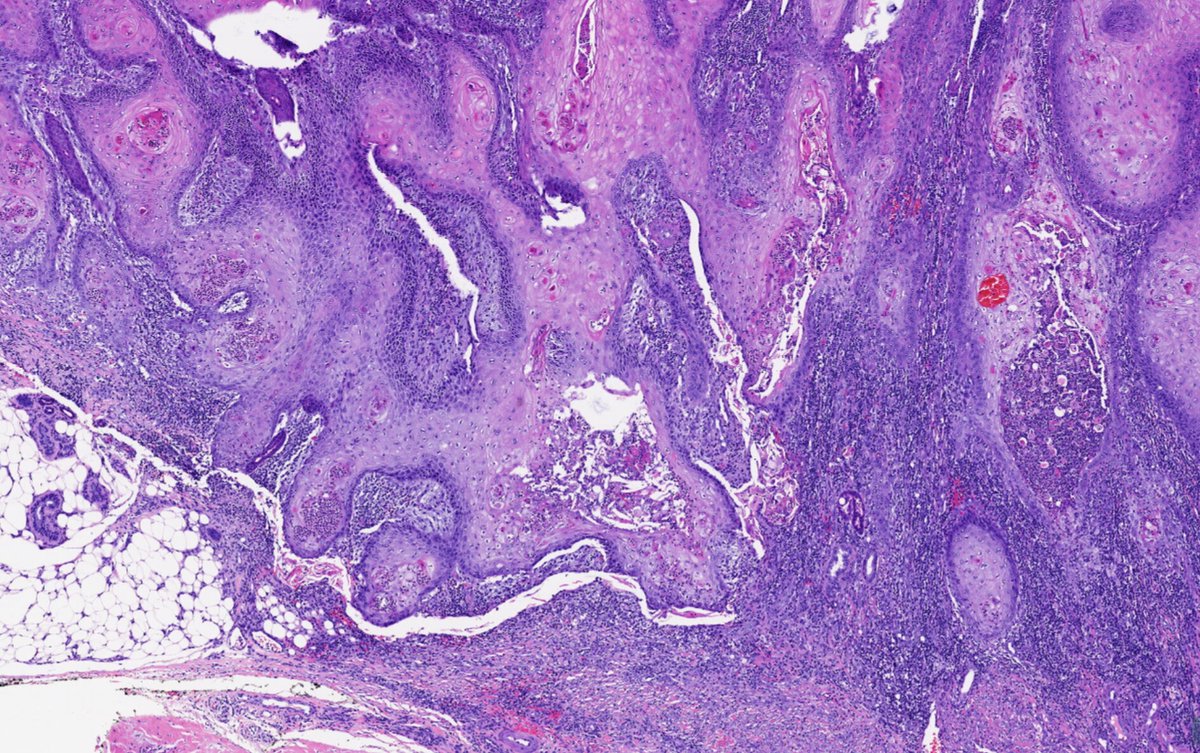
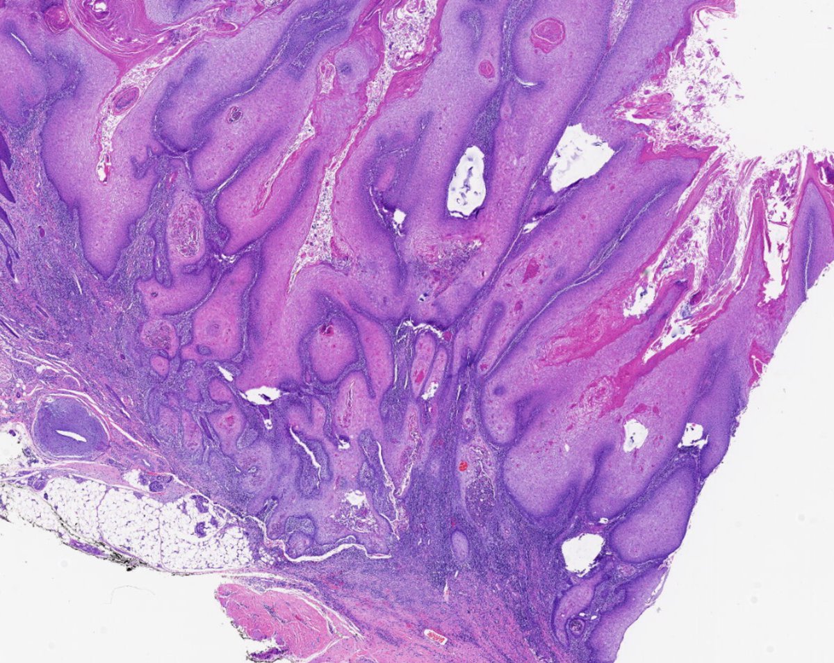
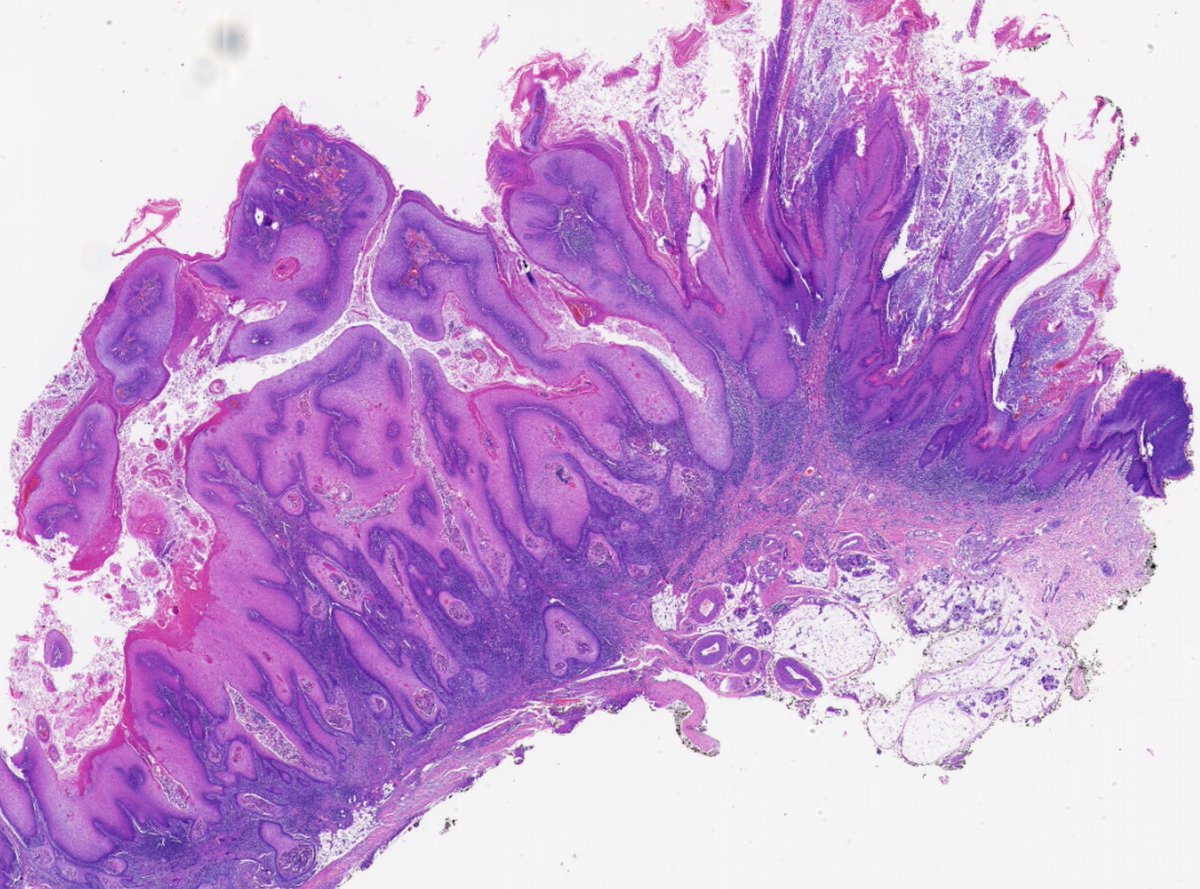
Verrucous squamous Lesion.
77 year old. hand. duration 10 years
#dermapth
Without @RACARR51 cocktail stains (P53, P16, Ki67), what would you call this lesion? I will not call it verrucous carcinoma for sure...@mccalmo @david_terrano @tonitatirado

Soft tissue lesion on a 70s man ring finger. Opinion appreciated.
#dermapth #sftpath
@DrGeeONE @DanGrahamMD @kis_lorand @pepeheffernan @MarcelaSaebL @tonitatirado @david_terrano @et565 @DiegoMoralesN @EssaoudiMohamed
More images follow:


In this case, it’s not at all difficult to distinguish between invasive melanoma and a dermal nevus. Just look at the difference in the nuclei size with SOX10 #dermatology #pathologist #dermapth #pathology #MedX #PathX #dermatologia


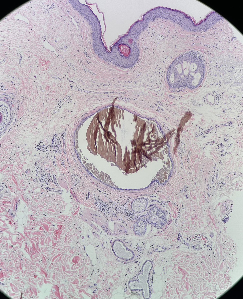
Cheek Lesion. Rule out melanoma. Due to this hidrocystoma content— what are those brownish materials? #dermapth

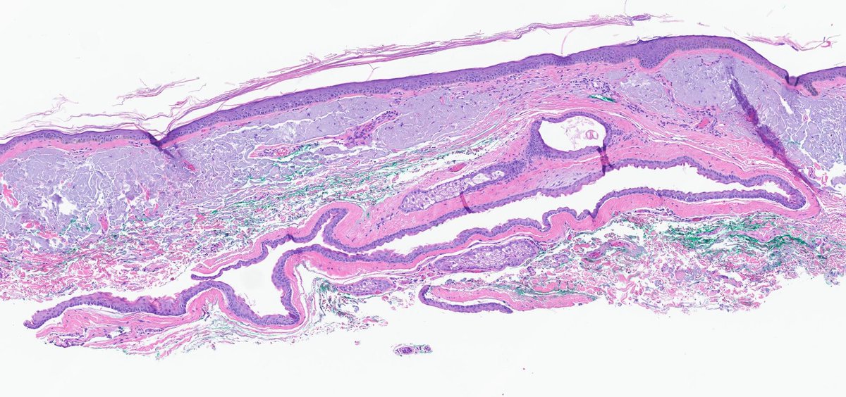


Xanthomatous infiltrates are hard without clinicals. So many different entities can have similar histopathological appearances. here is a lesion from 60s man arm (generalized infiltrative orange-yellow papules coalescing into annular plaques). #dermapth #xanthomatousinfiltrates
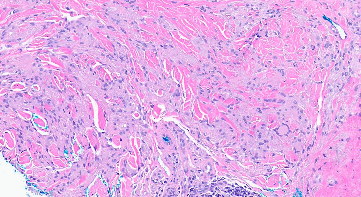


Intriguing #dermapth. what is this ? 14 year- old back
@DanGrahamMD @david_terrano @et565 @DCSkipperDO @MarcelaSaebL @redsnapperpath @ArjunRamaiya1 @mccalmo @kis_lorand @gonzadetoro @DrGeeONE @atman_ci @Histopatolomon


Be aware Trap! Not all collagen “trapping” is dermatofibroma —sclerosing hypopigmentated blue nevus can “trap” you too
#dermapth #dermresidents #PathTwitter #pathresidents
@VHNguyenMD @RashDecisionz @david_terrano @et565 @MarcelaSaebL @PIorfino @gonzadetoro @pepeheffernan


We are looking forward to working with @isabelcolmenero on #GermCellTumors and also learning from her on ##PediPath and #dermapth

Congratulations to Dr. @isabelcolmenero on receiving the McAdams Short-Term Study Stipend! #PediPath #SPPfall2022


Pseudoxanthoma Elasticum!
Did you heard about it? Me never! This is my learning today
#PseudoxanthomaElasticum free access guidelines https://t.co/EBX7Ea0zKm via @WileyHealth
The #dermapth conversation on this rare #skin disease https://t.co/8HdqZppqTt #dermtwitter #dermatology

1/
How would you call this pigmented lesion on the arm of 26yo/F patient? I thought this could be pigmented nevus of Reed. Should this pagetoid aspect of the lesion worry me? I would appreciate your comments on this one. #dermapth #dermatology #PathTwitter #melanocytic


woman in her 40s presented with a skin colored papule on her cheek. it's been growing in size for 6 months now. are you worried? #dermapth #dermatology #PathTwitter #melanocytic @melanopath @MELanocyteMD @Melanchocyte @PIorfino @MarcelaSaebL @redsnapperpath @et565 @DermpatGal

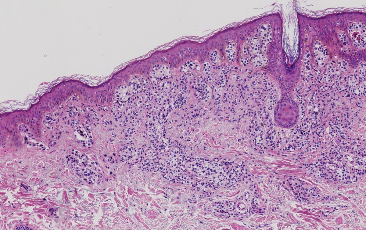


Woohooo!! It has been published!! Highly recommend this amazing read to all of you my friends ❤️



Most Popular Users

Elon Musk 
@elonmusk
240.5M followers

Barack Obama
@barackobama
118.7M followers

Cristiano Ronaldo
@cristiano
114.4M followers

Donald J. Trump
@realdonaldtrump
111.8M followers

Narendra Modi
@narendramodi
107M followers

Rihanna
@rihanna
98.7M followers

NASA
@nasa
92.2M followers

Justin Bieber
@justinbieber
91.8M followers

KATY PERRY
@katyperry
90M followers

Taylor Swift
@taylorswift13
84M followers

Lady Gaga
@ladygaga
75.5M followers

Virat Kohli
@imvkohli
73.4M followers

Kim Kardashian
@kimkardashian
70.8M followers

YouTube
@youtube
68.7M followers

Neymar Jr
@neymarjr
66.3M followers

Bill Gates
@billgates
65.2M followers

Selena Gomez
@selenagomez
63M followers

The Ellen Show
@theellenshow
62.1M followers

CNN
@cnn
61.7M followers

X
@x
60.6M followers