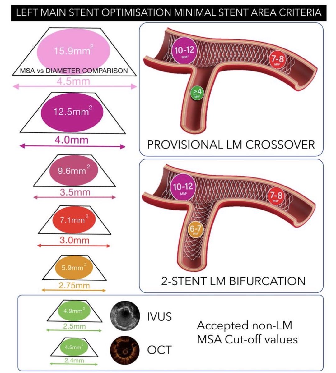
After 2 years of work, the 🇪🇺 consensus on #IVUS#OCT use in #LM#PCI is now published in #EHJ. Great international collaboration coordinated by #EBC and #EAPCI !!!
It includes fantastic practical images that can be hung on the walls of all cath labs…
https://t.co/2TbBTIoMxu
CAD with Atrial Fibrillation & RBBB
Is ECG Useful? Undoubtedly, yes. No instrument can replace it. However, for more than 120 years, the images recorded by conventional ECG have remained unchanged. 99% of them are high-frequency convoluted waveforms.
Coronary Artery Disease (CAD) was discovered in 1973 and officially named by Harvard in 1976. Yet in clinical practice, doctors face enormous challenges. AMI/CAD/ACS accounts for more than 60% of cardiovascular diseases — even higher in some countries. Doctors frequently encounter cases where the ECG clearly shows a CRBBB pattern, but the final diagnosis is CAD, with coronary angiography (CAG) revealing stenosis or occlusion greater than 70%. The issue is not that the ECG image is poor, but that its resolution has been insufficient for over 120 years.
The new scientific discipline of “signal processing” — a key technology of the Third Industrial Revolution — only developed after the year 2000. Unfortunately, it has not been applied to traditional ECG image processing.
Traditional ECGs do capture all the signals through convolution, but unfortunately, the fine details are not displayed. It is similar to how X-ray and CT images of the same patient contain the same natural anatomical elements, yet reveal different diagnostic information depending on the modality. The same principle applies to traditional ECG versus new-generation ECG — the difference lies in the level of image detail.
Please refer to the attached Figure showing ECG images of 4 patients taken before their CAG results.
PhysioSign USA
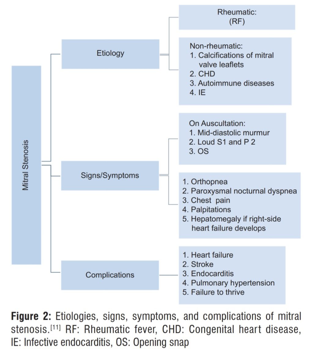
Update: Estenosis Mitral. 🫀💥
📄Revisión con repaso y comparación de directrices actuales ACC/AHA y ESC. 💯👌🏻
⬛️La EM sigue siendo una causa relevante de valvulopatía a nivel global: reumática en muchos países y calcificada en adultos mayores en contextos de altos ingresos. Se asocia a FA, tromboembolismo y peor pronóstico cuando es sintomática.
🔎Diagnóstico y cuantificación:
✔️ La ecocardiografía es la base (TTE; TEE para trombos en AI y mejor definición anatómica).
✔️Enfoque multiparamétrico: área valvular, gradiente medio y presión pulmonar.
✔️3D mejora la medición del área.
✔️Si hay discordancia entre síntomas y eco en reposo➡️ prueba de esfuerzo o evaluación hemodinámica invasiva.
🕵♂️ Seguimiento:
✔️La EM progresa lento, pero el eco seriado es clave incluso en asintomáticos.
✔️Descompensan con FA de novo, embarazo, anemia, fiebre o taquicardia.
💊 Tratamiento médico:
✔️Anticoagulación si FA, trombo previo o trombo en AI.
✔️Control de FC si taquicardia.
✔️Diuréticos para congestión (puente a intervención, no definitivo).
🛠️ Tratamiento definitivo (según anatomía y clínica):
✔️Valvuloplastia mitral percutánea: primera línea en EM reumática con anatomía favorable (score de Wilkins bajo <8 puntos), sin trombo en AI ni IM moderada-severa.
✔️Cirugía (comisurotomía o reemplazo) si no es candidato a percutáneo o falla del procedimiento.
✔️En EM calcificada del adulto mayor: intervención solo en casos muy sintomáticos y seleccionados (alto riesgo).
📄🆓️⤵️
DOI: 10.4103/ACCJ.ACCJ_24_24
https://t.co/0JZVDPCXuO
Comparative Effectiveness and Outcomes of Nebivolol Versus Other Beta Blockers in Patients With Hypertension: A Multicenter Cohort Study
In this retrospective cohort study of patients with hypertension without heart failure, nebivolol was associated with greater reductions in blood pressure and heart rate, as well as a lower risk of all‐cause mortality and major adverse cardiovascular events, compared with carvedilol and other beta blockers.
#Cardiology #MedTwitter #CardioTwitter #HeartHealth #Healthcare
@CMichaelGibson@DrMarthaGulati@hvanspall@AndrewJSauer@Hragy@AnastasiaSMihai@biljana_parapid
https://t.co/AqKmpwwTGi
In this infographic you have a structured synthesis of current screening recommendations based on official guidance from:
➡️ United States: U.S. Preventive Services Task Force (USPSTF), American Cancer Society (ACS).
➡️ Europe: European Commission (EU Council Recommendations 2022 update), and position statements from European Society for Medical Oncology (ESMO).
➡️ Only population-based screening with sufficient evidence is included.
Evaluación ecocardiográfica de la función diastólica del VI en el adulto. 📄Diagnostics 2026. 💯
🟠La valoración diastólica del VI sigue siendo un pilar de la ecocardiografía integral. No solo estima presiones de llenado, sino que es clave en el estudio de la IC FEVI preservada.
🔎 Enfoque actual multiparamétrico➡️ No existe un solo parámetro definitivo. La recomendación es integrar mediciones “clásicas” + técnicas avanzadas, adaptadas al contexto clínico y comorbilidades. 🤔⤵️
🟠Parámetros primarios:
▪️Flujo mitral (E, A, E/A, DT).
▪️Doppler tisular (e’, E/e’).
▪️Flujo venoso pulmonar (S, D, ar).
▪️TR e IVRT.
▪️Volumen auricular izquierdo indexado (LAVi).
▪️GLS del VI.
⚠️Disfunción si: E/e’ >14, LAVi >34 ml/m², TR elevada (>2.8 m/s) y e’ reducido orientan a ⤴️presiones de llenado.
🟠Técnicas avanzadas:
▪️Strain auricular (LARS)➡️ fuerte valor pronóstico en FE preservada.
▪️Myocardial Work (MW)➡️ puede aportar información indirecta sobre disfunción diastólica.
▪️Time-Harmonic Elastography (THE)➡️ nueva herramienta para rigidez miocárdica.
⚠️Limitaciones relevantes: edad, carga, arritmias, ventana acústica, variabilidad inter-vendor y dependencia del operador siguen siendo retos importantes.
📄🆓️⤵️
https://t.co/L3f77M9oeQ
https://t.co/DgkMutQpGe
Cardio pearls you should never miss
Part- 2
🔴 1️⃣1️⃣ ST elevation in aVR + diffuse ST depression
Think Left Main / proximal LAD / severe triple vessel disease
• ST depression ≥1 mm in ≥6 leads
• ST elevation in aVR ≥ V1 strongly suggests LMCA
• Widespread horizontal/downsloping ST depression
• Often hemodynamic instability
• Represents global subendocardial ischemia
• High mortality pattern — activate cath lab
Not “just NSTEMI.”
🔴 1️⃣2️⃣ Tall R in V1 + ST depression V1–V3
Think Posterior MI (posterior STEMI equivalent)
• R/S ratio >1 in V1
• Upright T waves in V1–V3
• Horizontal ST depression
• Mirror image of posterior STE
• Confirm with V7–V9 (≥0.5 mm STE)
• Often associated with inferior or lateral MI
Hidden STEMI.
🔴 1️⃣3️⃣ Regular narrow tachycardia ~150 bpm
Think 2:1 Atrial Flutter until proven otherwise
• Ventricular rate ~150 (atrial ~300)
• Sawtooth flutter waves in II, III, aVF
• Flutter waves may be buried in QRS/T
• Vagal maneuvers or adenosine unmask flutter waves
• Pseudo r’ in V1 may be seen
Don’t label as “SVT” casually.
🔴 1️⃣4️⃣ Irregular wide QRS tachycardia
Think AF with WPW until proven otherwise
• Very fast rates (>200 bpm)
• Beat-to-beat QRS morphology variation
• No consistent P waves
• Delta waves may be visible in sinus rhythm history
• Avoid AV nodal blockers (beta blockers, CCB, digoxin, adenosine)
• Use procainamide or cardiovert
High VF risk.
🔴 1️⃣5️⃣ Coved ST elevation V1–V3 + syncope
Think Brugada pattern (Type 1)
• ≥2 mm coved STE in V1–V2
• T wave inversion following STE
• Incomplete/complete RBBB morphology
• Often normal echo
• Fever can unmask pattern
• Young patient + syncope = high sudden death risk
Not “early repolarization.”
#MedTwitter #MedX #cardiology
@DrAkhilX @albertoortegana@dr_manish_ydv@daoo100@SameerYogi14@DocPriyamMD@shakilED@MiguelP23970914@DrYdv_hitesh01@TrackYourHeart@MedLearnHub@Frances98392343
👇 Overview of Double firing on ECG — also known as Dual AV Nodal Non-Reentrant Tachycardia (DAVNNT) 👇
👉 Mechanism: Simultaneous antegrade conduction over fast and slow AV nodal pathways, producing the classic pattern of one P wave followed by two QRS complexes.
➡️ Frequently misinterpreted as PVCs, PACs, or even atrial or ventricular tachycardia
➡️ Misdiagnosis may lead to unnecessary antiarrhythmic therapy
➡️ Patients can present with palpitations, dizziness, or presyncope
👉 EPS is key to confirming dual AV nodal physiology and distinguishing it from other arrhythmias (para-Hisian PVCs)
👉 For symptomatic patients, RF catheter ablation targeting the slow pathway is the treatment of choice, with high success and low complication rates, consistent with ACC/AHA/ESC recommendations.
💡 #EPeeps 💡
Awareness of this entity is essential to ensure accurate diagnosis, appropriate management, and avoidance of overtreatment.
🙏🙏 Another excellent video—brilliantly created by @N_Trajkovska , as always. 🫡🫡
@Phiso_de@MBraunEP@MoneebKhalaph@Cardioschool@chris_sohns@Ecgloverr@ECGEPSCADEVICE@ecgandrhythmRoe@ecgrhythms@makkyecg@KostekMilan@jjmt_ep@AbbottNews #CardioTwitter @AGEP_DGK@YoungDgk@ArashArya_EP@TobiasReichlin #LearnEP @Koichi16423232@MattersoftheH14@alex1708ander@DrRajeshG1
📘 Aneurisma del Septum Interauricular
Dilatación localizada del septum (usualmente en la fosa oval) que protruye hacia una o ambas aurículas, con movimiento excesivo.
Puede asociarse a FOP y eventos embólicos.
#SONECOM#GlosarioSONECOM#Ecocardiografía
🫀⚠️ No todo PVM es benigno
Existe un fenotipo con riesgo arrítmico. El PVM se define como protrusión sistólica ≥2 mm de las valvas mitrales en PLAX.
👉 Más en: https://t.co/M3KA6UxtGp
#SONECOM#Eco360#ProlapsoMitral#Ecocardiografía
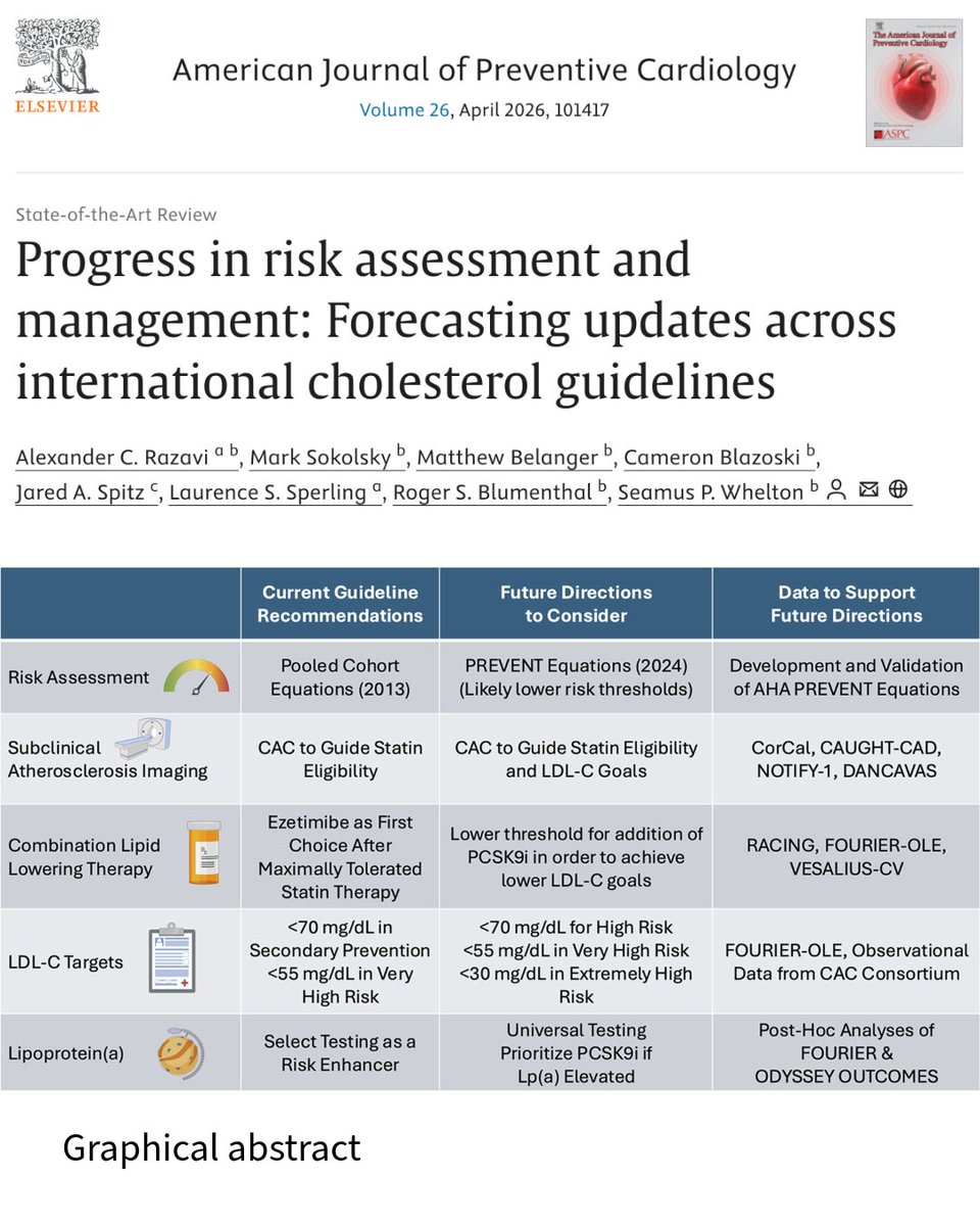
New advances in risk assessment and management are shaping updates across international cholesterol guidelines. This forecast highlights shifts in interpretation, thresholds, and treatment strategies to better prevent ASCVD worldwide.
🔗 https://t.co/3g50mJAcBB
#Cholesterol