Excited to share our new paper on elucidating the tRNA capture mechanism of the T-box mRNA. We use a combination of 2 & 3-color smFRET & different biophysical methods to unravel the conformational dynamics & directly visualize the capture of tRNA by mRNA
https://t.co/e7DPXnD36M
In a medical milestone, a customized base editor was developed, characterized in human and mouse cells, tested in mice, studied for safety in non-human primates, cleared by @US_FDA for clinical trial use, manufactured as a complex with an LNP, and dosed into a baby with a severe, rapidly progressing genetic disease... all in an astounding 7 months. Best of all, the infant patient shows apparent benefit. Congratulations to @kiranmusunuru, Rebecca Ahrens-Nicklas, and other team members for this heroic and inspiring effort, which has implications for the hundreds of millions of patients that suffer from thousands of genetic diseases.
https://t.co/wsgvvRYPVM
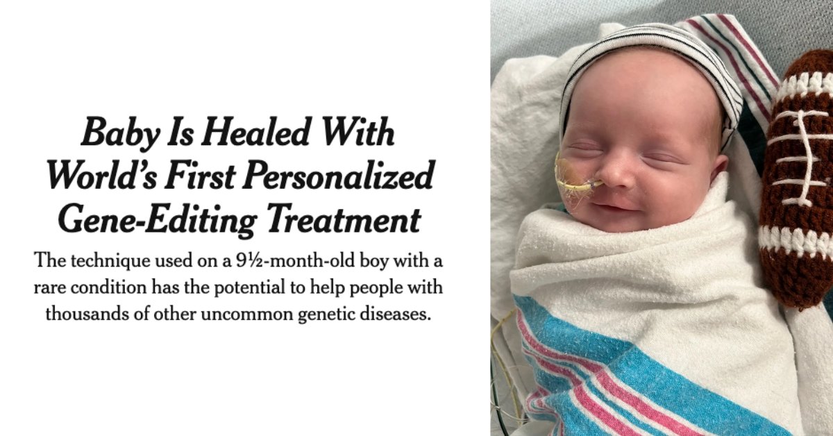
A baby named KJ, who was born with a rare genetic disorder – CPS1 deficiency – is healed thanks to doctors at the @ChildrensPhila who administered the world’s first personalized gene-editing treatment. The research team used base editing, a gene-editing method invented by HHMI Investigator @davidrliu and his lab at @Harvard University in 2016.
Earlier this year, Liu received the @brkthroughprize for developing both base editing and prime editing. These gene-editing technologies enable the correction or replacement of virtually any genetic mutation, including those that cause countless human genetic diseases.
Read more about Liu's work here: https://t.co/NlE25O7wJR
@jinweizhang@CharlesBNader J2 antibody is indeed the gold standard in RNA therapeutics for dsRNA detection. Congratulations Charles, Jinwei and all the authors for this fantastic work!
How to find fantastic new #RNAs? The J2 antibody is the gold standard to detect, map & enrich dsRNAs. Co-led by @CharlesBNader, Kevin Juma, Ankur Bothra & Steve Leppla’s lab, we visualize how J2 recognizes dsRNA and define its epitope, specificity & bias. https://t.co/ELXCvLv4ap.
How sturdy are base pairs near the ends of #RNA hairpins? Perhaps not as stable as one might think, esp. when there are bulges near and far. Led by @CharlesBNader in @NatureComms, we describe complete structures of #HIV#TAR RNA and its dynamic excursions.https://t.co/2vvKK1CQaC
I am very happy to announce that we solved the in-virus #cryoEM structure of the #HIV matrix protein. The structure helped us to resolve a long mystery in HIV biology. A🧵1/5
Read more in @Nature : https://t.co/rVDC5LomAn 🧪
Biological systems find ingenuous solutions. Almost anything that can happen, happens.
Still, I would not have guessed a role of RNA-binding proteins on the cell surface.
The Flynn lab are boldly investigating glycoRNAs and following their leads.
If u think this work is cool, note that it was done by grad students, PDs and staff in academic labs funded by federal tax payer dollars. If no federal funding, a lot less of this kind of research. Use your voice to protect federally funded research which is under attack. Thank u
Congrats to the @mammothbiosci team on achieving efficient muscle editing with the ultracompact #CRISPR nuclease NanoCas, an important breakthrough that paves the way for curing a wide range of genetic diseases! A nice overview from @ScienceMagazine here:
https://t.co/XQSjt5fq1l
Excited to share my recent article in @the_hindu for their Building Block series! I explored an important public health issue related to snakebite & antivenom accessibility in India.
Why are antivenoms not easily accessible in India? - The Hindu https://t.co/2Ic383sizA
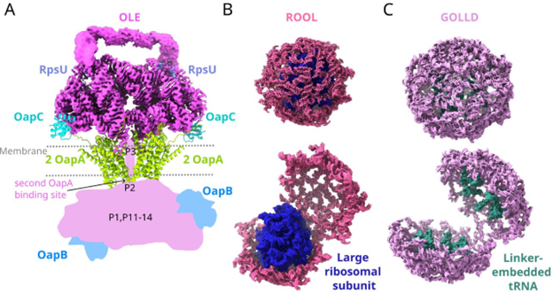
#ncRNA molecules are stranger than you think. We propose that OLE RNA is bacteria's functional mimic of
#mTOR. The Das lab at Stanford proposes OLE RNA penetrates cell membranes! They also find that RNAs we called ROOL and GOLLD form 8-membered nanocages. https://t.co/DQ7Zr5p6DT
@RachaelKretsch@RDasLab@chiulab Wow, these RNA structures are amazingly beautiful!! Always wondered what is the role of these long non-coding RNAs in bacteria!! Excellent work, Congratulations to all the authors! 🎉👏🏼
Our latest work is out in @Nature today! Using smFRET, we directly visualized recruitment of the eIF4F complex to the 5' cap of eukaryotic mRNAs and formation of an activated mRNA. Our findings reveal new and surprising roles for each eIF4F component. 1/3 https://t.co/THosKn33fC
I’m proud to share the exciting news that all undergraduate students from households earning $75,000 or less will be able to attend any UMass campus tuition-free starting next fall: https://t.co/6JHESltrlR @umassamherst@umassboston@umassd@umasslowell#umass#highered (1/4)
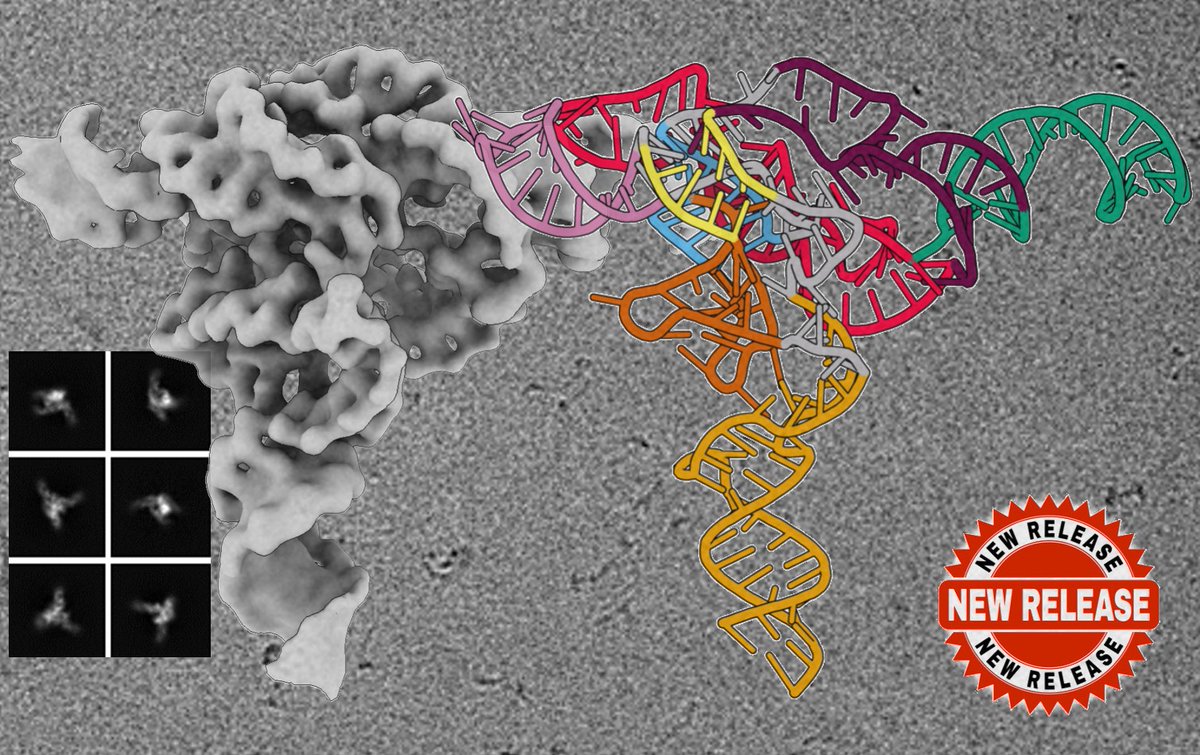
@3DRNA_CryoEM:
Cryo-EM structure of raiA ncRNA from Clostridium reveals a new RNA 3D fold
https://t.co/ftR6Psd8cV
@JMolBiol
Thanks @SOLARIS_science for the great microscope facility.
3D model illustration made with @proteinimaging