How wonderful to have our very own @loft_lab featured on @AcademicMinute today!
Give it a listen and learn more about his new 🧠 imaging technique, which opens new possibilities for detecting early signs of vascular and neurodegenerative disease.
https://t.co/TNFBm87hO0
High resolution iso-1mm 7T ASL allows assessing molecular, cellular and transcriptomic bases of laminar perfusion and cytoarchitecture coupling in the human cortex https://t.co/V0tz2Ioob8
Happy to share our analysis results of ADNI-3 ASL data, showing consistent trend of CBF decrease with AD progression in key AD related brain regions in both GE and Siemens ASL data https://t.co/NvVcUl6oFv
Saturday’s CAMERA–OSCMRM session “Low-Cost Open Source MRI: Are We There Yet?” at #ESMRMB2025 brought diverse insights on open-source MRI — from hardware development to radiology and education.
Thanks to our speakers for thoughtful talks and discussion! #LowFieldMRI#OpenSource
Here's a reminder on #InternationalCoffeeDay: It's OK to take a (coffee) break. ☕
"It’s ... helpful to have a venue to share the day-to-day ups and downs of life as a grad student," a #PhD student wrote in this 2019 #ScienceWorkingLife. https://t.co/L9cA1BH4qi
Glad to share our new paper in Nature Cardiovascular Research https://t.co/14UFbALL5O introducing a new method for mapping microvascular pulsatility using high resolution VASO and ASL MRI at 7T @layerfMRI
🚨 New discovery 🚨
Our @loft_lab developed a noninvasive brain-imaging method that reveals hidden microvascular pulsations in aging brains, which may offer earlier clues into dementia and Alzheimer’s risk.
Read the press release here: https://t.co/gWhak1o15z
A single aquaporin protein lets one billion water molecules into a cell each second. It is one of the "fastest" channels in all of biology.
Each water flows through in single file, while H+, sodium, potassium, and other ions are excluded.
What explains this remarkable selectivity? Two clever protein filters.
But first: Each aquaporin protein is made from six alpha-helices that run back and forth through the cell membrane. These helices form an hourglass shape so that the channel is cone-shaped at either end and extremely narrow in the middle.
The two ends of the protein also mirror each other, with their point of symmetry occurring at the narrowest part of the channel. It's thought that modern aquaporins evolved through a gene duplication event.
Now let's zoom in a bit and look at the two filters within aquaporin that account for its extreme selectivity. Water can flow either way through the channel, so we'll imagine that we're moving from OUTSIDE the cell to INSIDE the cell.
The first filter we encounter is called the ar/R filter. This is a cluster of polar and charged amino acids that point inside the aquaporin channel.
These four amino acids are *usually* arginine (+ charge), histidine (polar), and two bulky amino acids like phenylalanine and tryptophan.
These four amino acids come together to restrict the middle of the pore, making it just wide enough for a single water molecule to pass. The positively charged arginine also helps "stabilize" water molecules while forcing away sodium ions or protons.
The second filter encountered on our journey into the cell is called the NPA motif (short for asparagine–proline–alanine). There are two of them inside each aquaporin.
These two protein motifs point at each other in the center of the aquaporin. As a water molecule passes the ar/R filter and moves deeper into the channel, it forms hydrogen bonds with the asparagine amino acid in the first NPA motif.
But because the two motifs are oriented in opposite directions, the local electric field at the channel’s midpoint next forces each water molecule to rotate 180° and latch onto the asparagine in the second NPA motif. In other words, each water molecule gets flipped around inside the aquaporin channel.
This "flipping" is important because it breaks the water molecules free from any hitch-hiking protons. It acts as the final filter.
My first book with @ShaoXingfeng on Imaging BBB Dysfunction in Brain Disorders is online for pre-order, it summarizes BBB biology, foundations and clinical uses of cutting edge imaging technologies for mapping BBB function in a healthy and diseased brain
📢 Now available for pre-order: Imaging Blood-Brain Barrier Dysfunction in Brain Disorders by our very own @loft_lab & @ShaoXingfeng!
This new volume explores the blood-brain barrier’s critical role in brain health and disease 📚🧠
https://t.co/2bTZ9VJDmf
Focused ultrasound with microbubbles can safely open BBB and the effect can be detected using DP pCASL MRI without contrast agent. Checkout the video and our preprint https://t.co/9uuSOX2SvT
📽️ New week, new animation with data from @loft_lab!
Focused ultrasound with microbubbles can temporarily open the blood-brain barrier, enabling noninvasive, targeted drug delivery while minimizing risk 🧠
Stay tuned for something big coming out of @loft_lab 👀
@mikolaj_pawlak BBB Kw changes were observed between 45 and 90 minutes after Focused Ultrasound -BBB Opening. We used a 2-minute focused ultrasound protocol combined with microbubbles for the treatment.
Focused ultrasound induced BBB opening in the Dorsal Striatum of Non-Human Primates can be detected by BBB water exchange imaging using DP pCASL https://t.co/9uuSOX2SvT
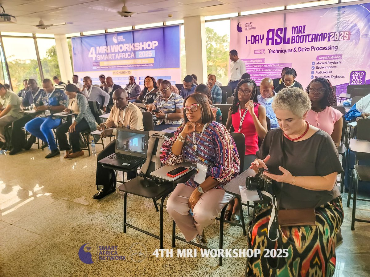
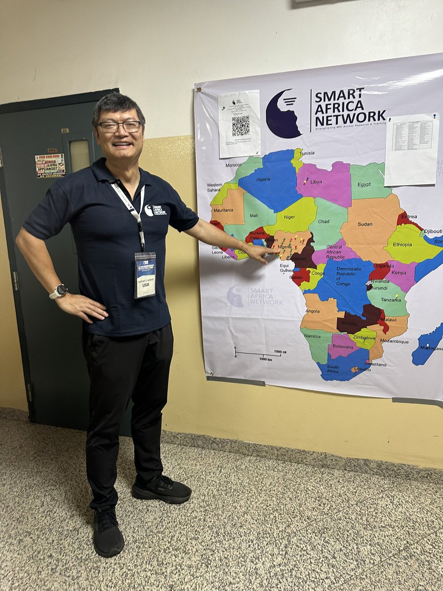
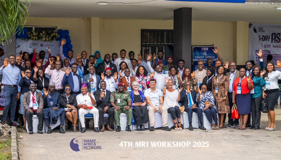
Honored to attend SMART Africa MRI workshop focusing on ASL in Lagos Nigeria 🇳🇬 impressed by the growth and passion of MRI in Africa, glad to share ASL experience and contribute to global MRI, congrats to @SMARTA_NETWORK
The 3rd annual meeting of 7T Translational Alliance of North America (7TANA) was successfully held at UTSW July 14-15 2025, great progresses in multiple frontiers of 7T clinical translations. Congrats to organizers Dr. Anke Henning and Sheeba Anteraper at UTSW see y'all next year
🌟 New week, new animation 🧠
Research by our @loft_lab uses noninvasive ASL MRI to show that brain blood flow decreases even in the earliest stages of Alzheimer’s — before memory problems appear. This could help doctors catch the disease earlier and track treatment over time.
Thanks for @audreypfan invite to visit UC Davis BME and imaging research center, impressive program great exchange with faculty and students, beautiful campus with an arboretum also thx @ydavidzhu