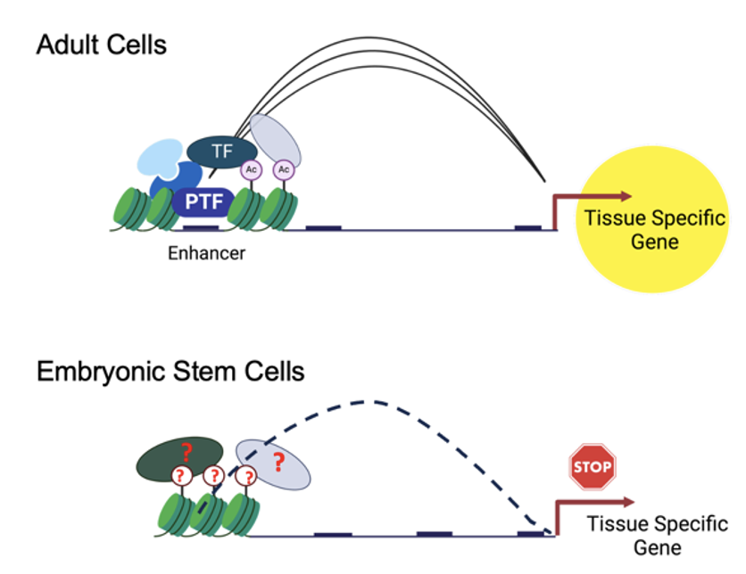
Are the enhancer regions that control the expression of lineage genes established from early embryonic stages or do they gain their transcriptional potential along differentiation? Today in @CellGenomics we address this question: https://t.co/atWlKbDUVq 1/15🧵
A fully iPS-cell-derived 3D model of the human blood–brain barrier for exploring neurovascular disease mechanisms and therapeutic interventions | Nature Neuroscience https://t.co/3GS4z2FjVo
Esta babosa marina puede desprenderse voluntariamente de su cabeza cuando está estresada o parasitada, y luego regenerar todo su cuerpo. Uno de los ejemplos más extremos de regeneración conocidos por la ciencia. 🐌🧬
✨Not a rainbow trout… a rainbow zebrafish 🌈 🐟
The expression of 10 different genes simultaneously including Hox genes along the body axis, somites, brain regions, skeletal muscle, and heart 🔬Video by Alice Sherrard, Gabby Jerz & Nipam Patel 🧪 #FluorescenceFriday
Finally here! Our preprint on the MOSAIC, a multimodal adaptive optical microscope enabling non‑invasive in‑vivo imaging from molecules to organisms, is out.
https://t.co/B2GHHP5b0K
@Eric_Betzig@legant_lab
One protein, two opposing roles: This ‘compelling’ study shows how the same signalling molecule can drive either protrusion or retraction in migrating cells depending on its concentration and activation dynamics.
https://t.co/iz3UrpXXQf
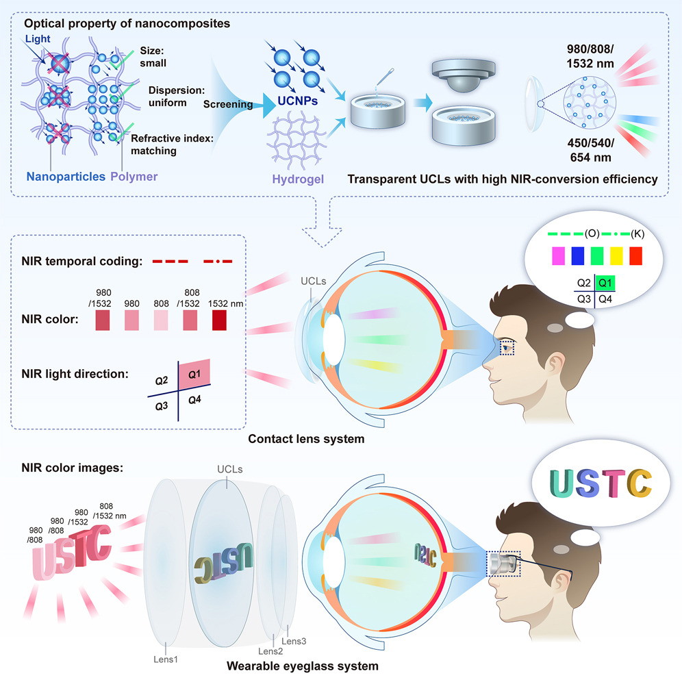
Infrared contact lenses allow people to see in the dark, even with their eyes closed. https://t.co/2Hgkq2ESF0
@USTCGlobal Yuqian Ma & colleagues
@CellCellPress
In which we discuss the homologies between amphibian and mammalian #organizers from the perspective of #gastruloids with special relevance to recently described A and C gastruloids as containing Spemann/Mangold and Trunk/Tail organizers https://t.co/AX9dWdUDEZ
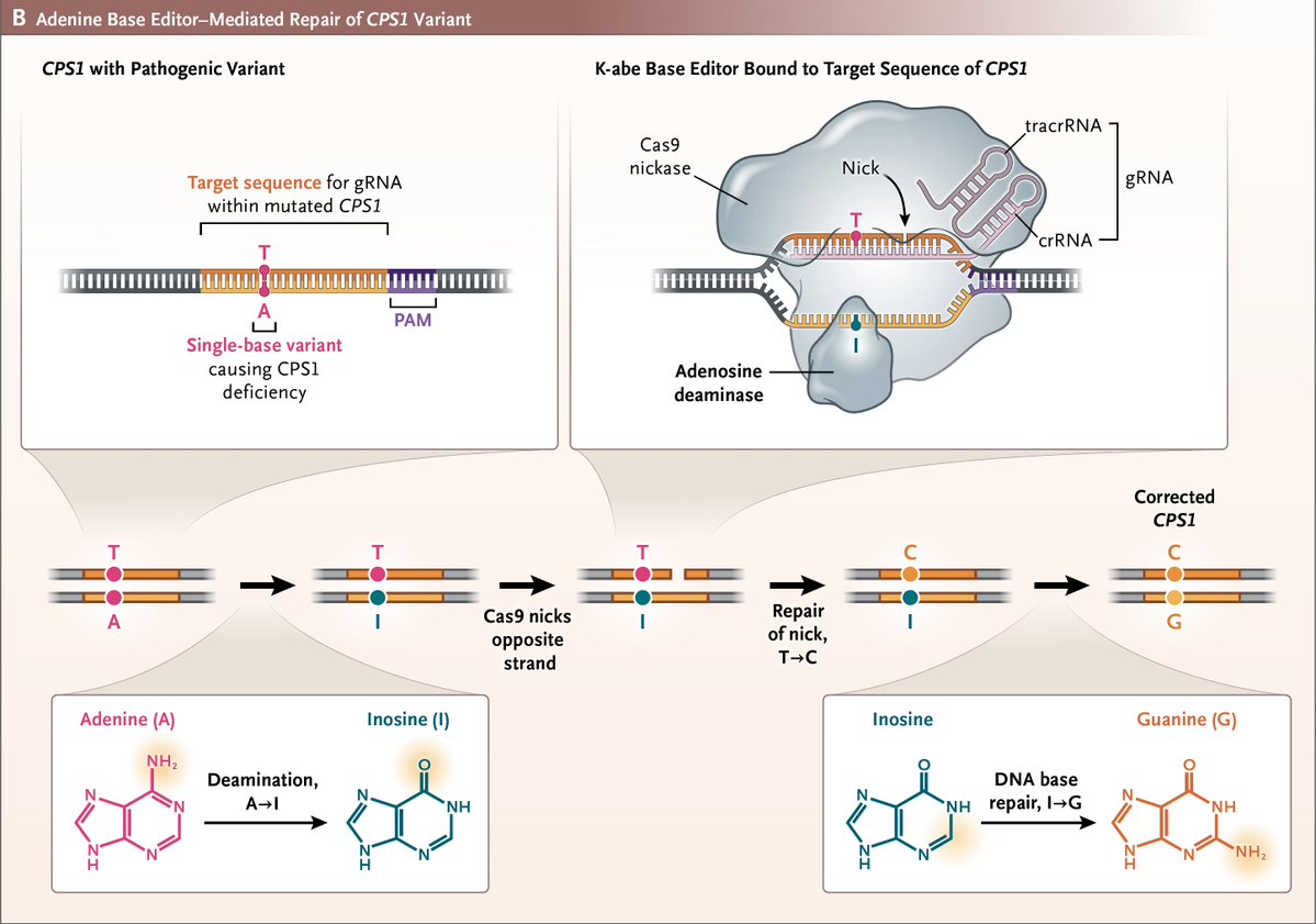
Today "a milestone in the evolution of personalized therapies for rare & ultra-rare inborn errors of metabolism"
—the 1st human to undergo custom genome editing
—outgrowth of decades of NIH funded research
https://t.co/HmJxYWDCV7
https://t.co/4DsNELnBut @NEJM
https://t.co/y7NMGPpXnE
Count your somites! To make a vertebra, it takes 120 minutes in mice, 90 minutes in chicks, 30 minutes in zebrafish, and ~4.5 hours in humans. #ZebrafishFunFacts
mScarlet3-H (aka mYongHong) makes a great pair with mBaoJin (https://t.co/mzs9doo1Ka) for long-term live cell imaging. All plasmids can be requested for free from WeKwikGene https://t.co/Aie1iHeuDw
This time-lapse captures 17h of axonal growth from a chick dorsal root ganglion explant, seen through the actin cytoskeleton using live imaging. I just submitted this video to the Nikon Small World in Motion competition. Today is the last day to upload yours!😉 #neuroscience
Coordinated contractions enable embryo elongation
Read this Research Highlight showcasing work from Flora Llense, Michel Labouesse and colleagues:
https://t.co/accuKOAGPC
🎥a pre-contraction embryo expressing Pmyo-3::GCaMP::GFP
Single-molecule localisation microscopy & live-cell imaging reveal fine details of chromosomal organisation underlying growth of E.coli
📷 @miCHRIScopy et al @Uni_WUE in @NatureComms
➡️ https://t.co/bH8CtbQAh5
Ever wonder what the architecture of a neural network would look like, in a novel organism that had not been through selection for specific structure and function of an embodied nervous system? Here's our #preprint with morphological, behavioral, electrophysiological, and transcriptomic analysis of a new kind of Xenobot with a nervous system:
https://t.co/CJLl8DR2JA - the hard work of @halehf@LaurieONeill99@mmsperry and @LPiolopez
Abstract: "A great deal is known about the formation and architecture of biological neural networks in animal models, which have arrived at their current structure-function relationship through evolution by natural selection. Little is known about the development of such structure-function relationships in a scenario where neurons are allowed to grow within evolutionarily-novel, motile bodies. Previous work showed that when a piece of ectodermal tissue is excised from Xenopus embryos and allowed to develop ex vivo, it will develop into a three-dimensional (3D) mucociliary organoid, and exhibits behaviors different from those observed in tadpoles of the same age. These 'biological robots' or 'biobots' are autonomous, self-powered, and able to move through aqueous environments. Here we report a novel type of biobot that is composed of ciliated epidermis and additionally incorporates neural tissue (neurobots). We show that neural precursor cells implanted within the Xenopus skin constructs develop into mature neurons and extend processes towards the outer surface of the bot as well as among each other. These self-organized neurobots show distinct external morphology, generate more complex patterns of spontaneous movements, and are differentially affected by neuroactive drugs compared to their non-neuronal counterparts. Calcium imaging experiments show that neurons within neurobots are indeed active. Transcriptomics analysis of the neurobots reveals increased variability of transcript profiles, expression of a plethora of genes relating to nervous system development and function, a shift toward more ancient genes, and up-regulation of neuronal genes implicated in visual perception."
Why do some neurons say “sleep” while others shout “stay awake!” — inside the same brain?
In fruit flies, researchers just mapped the molecular chaos behind this nightly tug-of-war. And it’s wilder than we thought.
https://t.co/hl8Npp3iJk