Annual killifish make neutrophils before almost every other cell type, even before gastrulation. Neutrophils usually arise from the lateral plate mesoderm (forming in cyan), but in killifish they are already patrolling around the embryo in gold. https://t.co/GeOzVd9H9M
New in @NatureMethods: a new human stem cell model of EMT lets us watch cells transition in both 2D and 3D and shows human stem cells can undergo the transition differently depending on how they are grown.
🔗 https://t.co/6Fpa2xJ4yc
I wrote about AI in academia. "PhD-level thinking", LLM bias, grunt work, alignment, AGI, data center water use, AI politics -- something for everyone.
What is the global structure of cell-state space—and how do perturbations drive transitions within it?
Excited to share our new preprint (https://t.co/ZTlAY4eaJf), a work in collaboration with @JswLab.
This group of French researchers (Sur le champ) trained about 200 re-enactors to test the crowd dynamics of hoplite warfare and routing more generally. It's really interesting how the often frustratingly vague statements of ancient writers become clear with this footage
Boston just got completely shut out of the 2026 James Beard Award nominees.
Not a single restaurant or chef from Massachusetts made the list.
The last time we had a major category win was in 2019.
Speaks volumes on the current state of Boston's restaurant scene this decade.
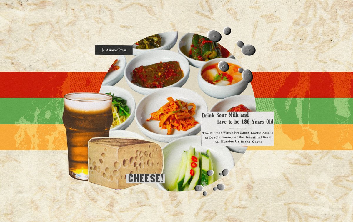
New Essay: CULTURE SHIFT
The human immune system is, in one sense, a detection mechanism. It has evolved, over millions of years, to scan the body for molecular signals that tell it whether to attack or stand down. Most of these signals come from pathogens, damaged cells, or the body’s own hormones. But in 2019, a lab in Germany published a finding that pointed to a much stranger source: one of the signals sensed by the immune system is found in sauerkraut.
When people eat sauerkraut, a molecule called phenyllactic acid (D-PLA) — found in fermented foods — enters their bloodstream and activates a receptor, known as HCA3, on immune cells, triggering an anti-inflammatory response. In addition to lactic acid, phenyllactic acid is one of many compounds produced by lactic acid bacteria during the fermentation of sauerkraut and related fermented foods. Prior to this study, other molecules had been found to bind HCA3, but D-PLA was a hundredfold more potent than any of them.
This discovery advances our understanding of how fermented foods can reduce inflammation, but more striking is what it suggests about hominid physiology. Although HCA3 is part of a larger family of receptors broadly conserved across eukaryotes, HCA3 is only present in humans and other great apes like chimpanzees and gorillas — and not even in other mammals. It is a recent addition to the genome, appearing only a few million years ago. Its existence seems to suggest that our immune system evolved to recognize the microbial metabolites from fermented foods.
We tend to think of fermented foods as something humans invented and then chose to eat. But, increasingly, scientific evidence suggests the causality runs the other way: Fermented foods appear to have helped shape human biology itself, and our bodies may have been built, in part, to expect them.
The case for this runs from changes in hominid gut anatomy millions of years ago to the HCA3 receptor, to a growing body of research linking fermented food consumption to immune function and gut health. And it raises an uncomfortable question about what happened when the Western food system, in the name of safety and efficiency, quietly removed these foods from our diets in the nineteenth and twentieth centuries.
Read the full essay: https://t.co/udEQu3YkUx
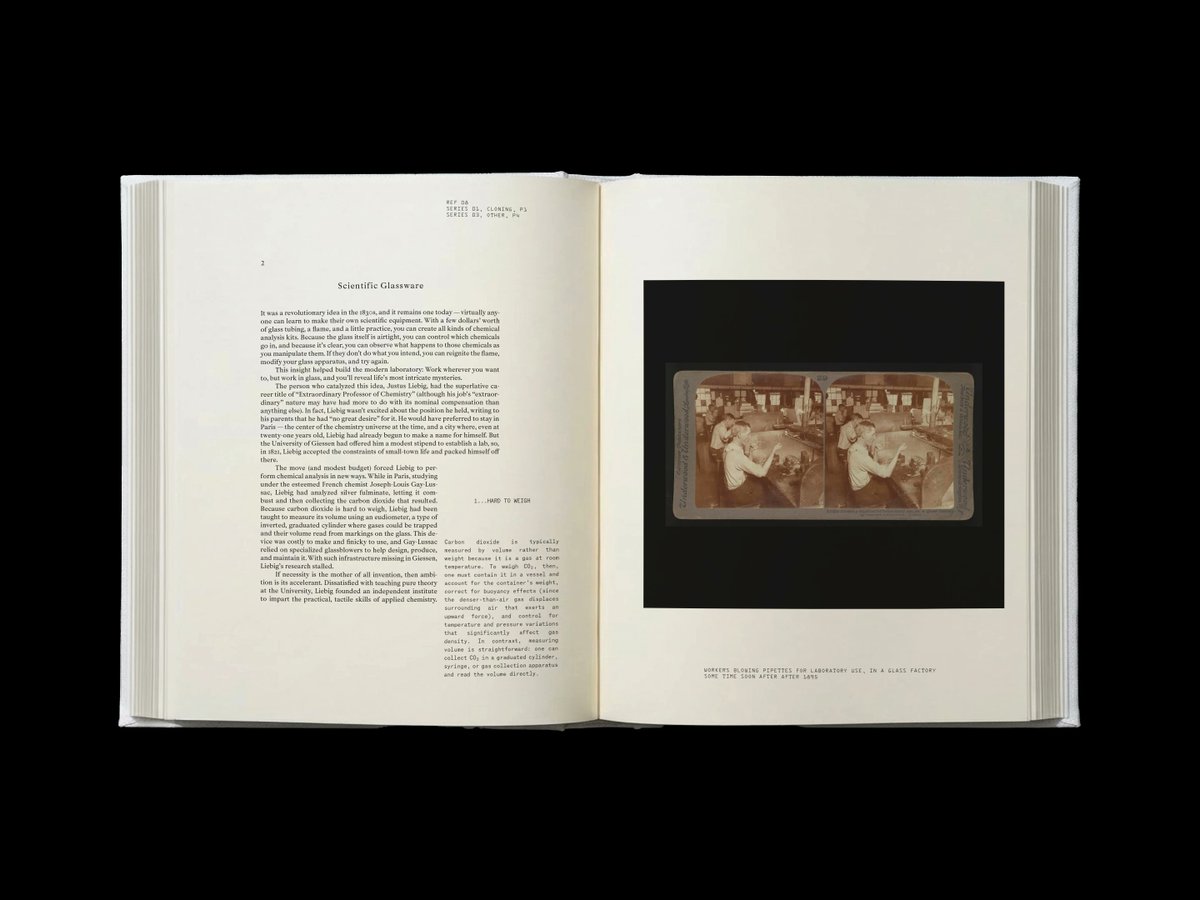
Have a photo from your research you'd like to see in print?
We’re collecting images for Making the Modern Lab, an upcoming book from Asimov Press about the tools, spaces, and experiments that define modern biology.
If your image is selected, we’ll pay $30 per photo and include a photo credit in the book.
Microscopy images, lab benches, instruments, experimental setups, and lab environments are all welcome. Phone photos are fine —we’re most interested in capturing the real look of modern research — but we are seeking well captured images with good composition. People are allowed in the photos, if the image clearly depicts the topic.
While this call is open, we have a list of images we are specifically searching for:
CHO cell microscope image
Liquid Handling Platforms (full view of instruments, action shots of liquid transfers)
A multiwell plate being fed into a plate reader.
Zebrafish (adult fish in tanks, microscope embryo images)
Biochemical buffers (labeled bottles lined up)
Bioreactors (any size)
C. elegans (full body or fluorescent microscopy. Ideal if neurobio related images)
Immunohistochemistry microscopy images
Electroporator setup
Liquid Chromatography setups (HPLC, HPLC-MS, or FPLC)
Mammalian tissue culture media
S. cerevisiae microscope image (ideal if you can get an image depicting cells in different parts of the life cycle.)
BONUS: Images of historical lab equipment in your lab.
Submit here: https://t.co/UHR2jzK2lv
Fascinating timelapse of an alpine newt (Ichthyosaura alpestris) growing from a single cell, into a complete, complex living organism over three weeks.
📽: Jan van Ijken
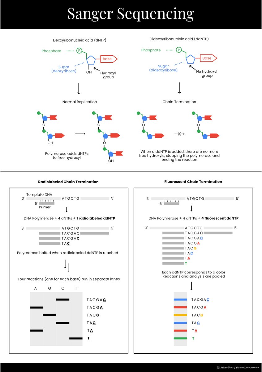
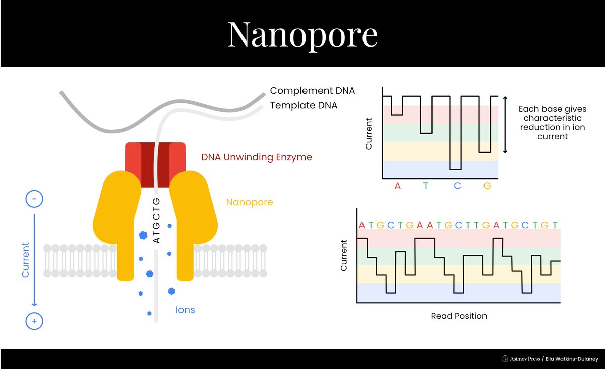
A Visual Guide to DNA Sequencing.
Learn how different DNA sequencing technologies work, from Sanger sequencing to Illumina to nanopores. (Complete with illustrations!)
Written by @evandeturk. Illustrated by @EllaWD_PhD.
Finally out! Patrick developed a system to study SET isoform specificity in ES cells, revealing a role for SETa/b switch in endoderm specification. Check it out🔥 @psl_lim https://t.co/jWgk7V8r0D
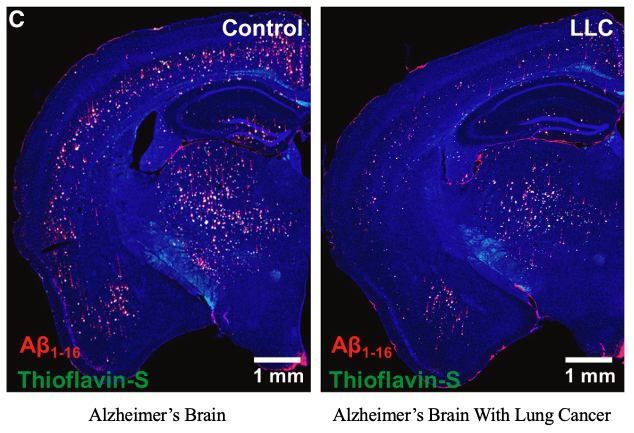
Cancer patients rarely get Alzheimer's.
And a 15-year study in @Cell just explained why. They found a protein that clears brain plaques in mice - by activating the brain's own IMMUNE CELLS 🧵
Thanks to the authors for sharing all components of this mouse embryo spatial transcriptomics data from cell gene counts to per-molecule coordinates: https://t.co/LVkzsL8GGT 🥳
So I vibe coded an app to explore the 3D subcellular transcript organization: https://t.co/GMiJ8ejZxt
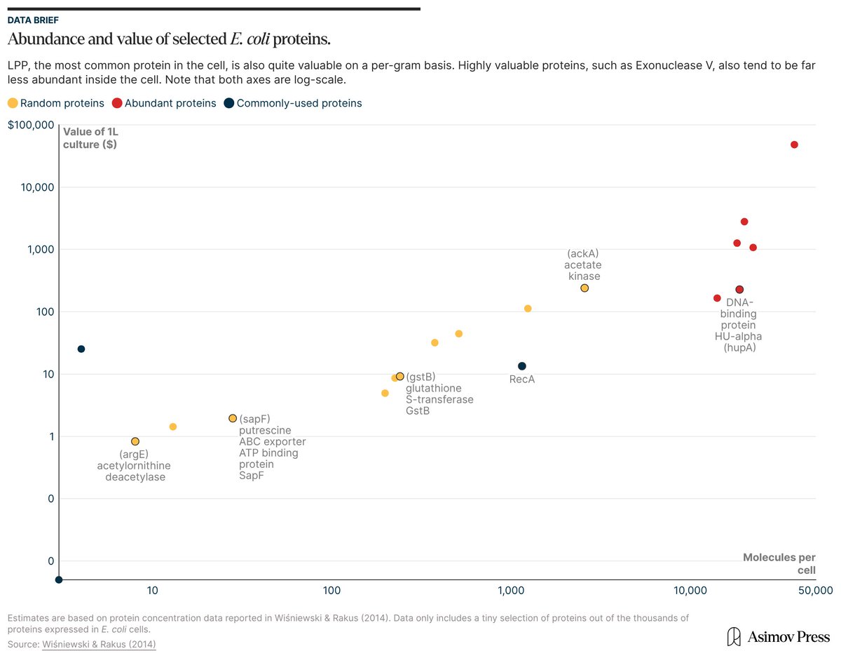
If you took a liter of E. coli cells, isolated all their molecules, and sold them, they'd be worth ~$600,000.
This is a thought experiment, of course; just doing the isolations might cost more than the molecules are worth.
But if you're keen to learn which parts of cells are worth the most money, check out our article: "The Price of E. coli."
https://t.co/QdbW8jofjg
When polishing scientific figures for publication, I try to limit fonts + font sizes to reduce visual noise. But doing this by hand is tedious.
So I vibe coded a web app to standardize text in SVGs: https://t.co/0JRhstRhAp
Try it out! Spot the differences. Hope it's helpful