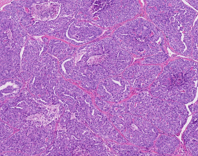
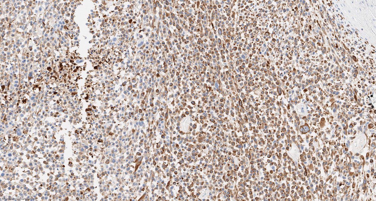
New in #HumPathol: L1CAM expression in eosinophilic solid and cystic renal cell carcinoma: clinicopathologic and immunophenotypic insights from a multicenter cohort. https://t.co/GpHZa4g5mC #pathology#PathTwitter#PathX#GUpath
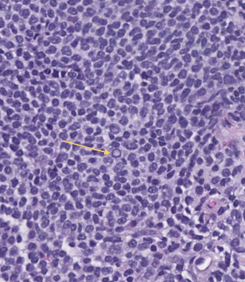
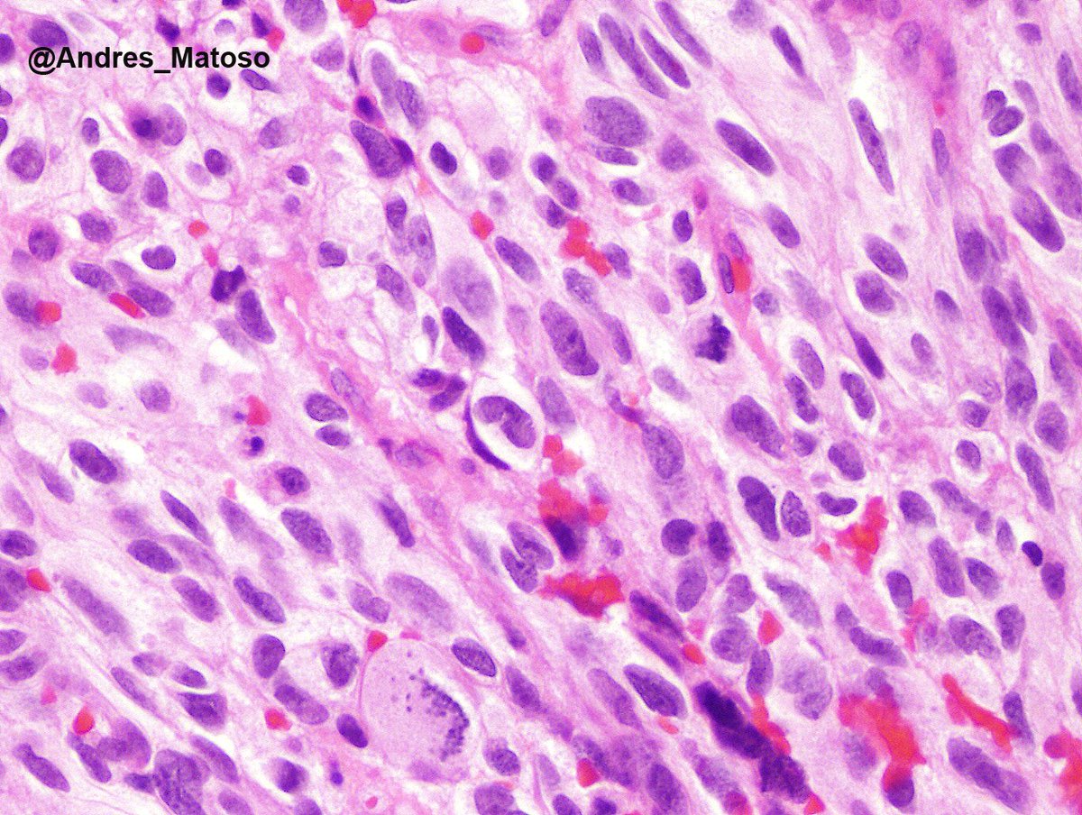
What's the diagnosis of this fourth ventricular tumor in an adult? The tumor has mutations in NF1, FGFR1, and PIK3CA. (It's a Rare, Generally Nonaggressive Thing.) #pathology#neuropath#pathtwitter
Atypical ductal hyperplasia (ADH) vs low-grade DCIS
Distinguishing them remains challenging, with significant interobserver variability among pathologists.
WHO recommends a conservative approach for lesions of limited extent, especially on core biopsy
The preferred quantitative criteria described by Dr. Page:
Low-grade DCIS requires complete involvement of at least two separate duct spaces by a proliferation showing both the cytologic and architectural features of low-grade DCIS.
ADH is typically small/focal (often <2–3 mm). Larger foci may still be accepted as ADH when associated with a radial scar/complex sclerosing lesion or papilloma.
• ADH → no need to report margins
• DCIS → margins required
Pic 2: Core biopsy, signed after consensus conference as multifocal ADH, approaching low grade DCIS.
Pic 3: Excisional biopsy/partial mastectomy: DCIS grade 1
Dr. Turner. Breast Pathology CAP on Demand https://t.co/flTFNvANfS
#pathology #CAPondemand #PathX #Breastpath #PathTwitter
Check out our case report:
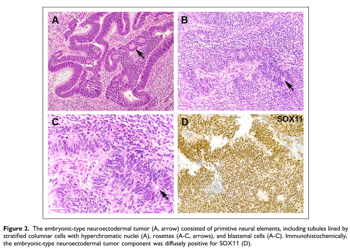
Testicular Mixed Germ Cell Tumor with Concurrent Embryonic-Type Neuroectodermal Tumor and Nephroblastoma
https://t.co/9ceFflpGjB
#GUpath
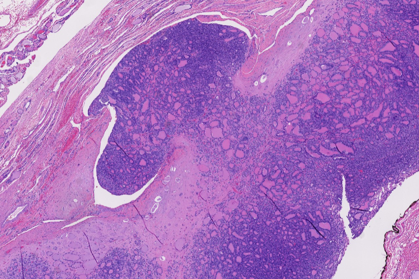
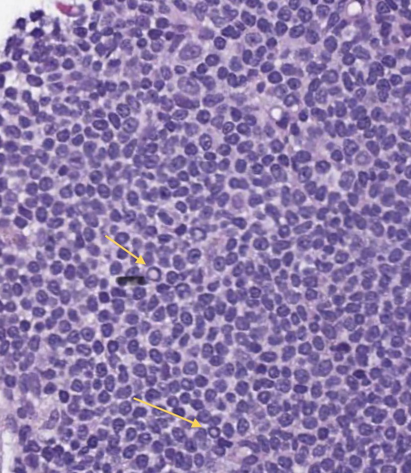
In the 2022 WHO Classification, poorly differentiated thyroid carcinoma (PDTC) is now grouped under high-grade follicular cell-derived non-anaplastic thyroid carcinoma (HGFCTC), alongside high-grade differentiated thyroid carcinoma (HGDTC)
Diagnosis of PDTC requires:
• Solid/trabecular/insular (STI) growth pattern
• Absence of PTC nuclear features
• ≥1 high-grade feature: mitosis >3/10 HPFs, necrosis, or convoluted (“raisin-like”) nuclei
Encapsulated poorly differentiated thyroid carcinoma (PDTC) with no invasion or limited invasion (capsular invasion or focal VI) has a more favorable outcome than widely invasive PDTC
These tumors should be subclassified as:
• Encapsulated with capsular invasion only
• Encapsulated angioinvasive
• Widely invasive
Always report encapsulation and extent of invasion. It may influence clinical management
Dr. Scognamiglio #USCAP #PathX #PathTwitter
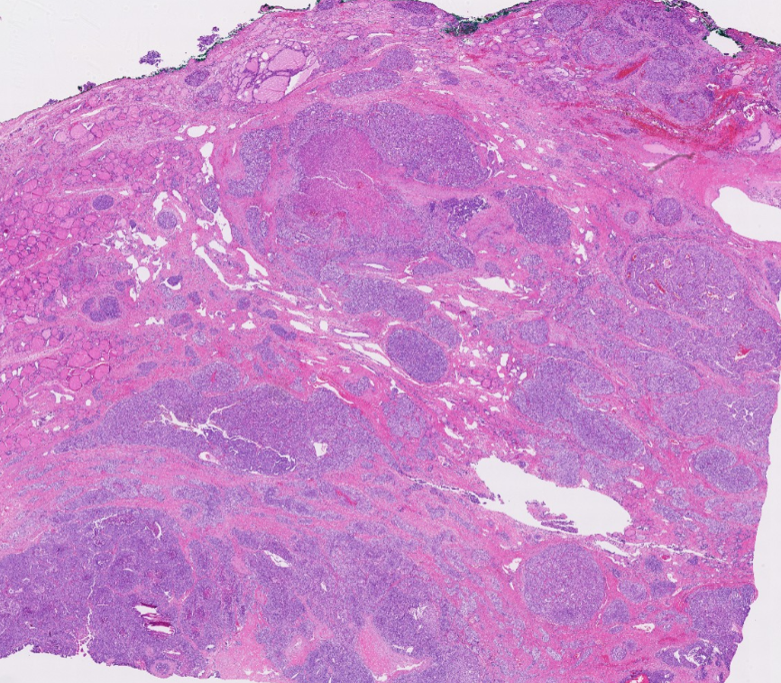
In the WHO 5th edition (2022), follicular thyroid carcinoma (FTC), invasive encapsulated follicular variant of papillary thyroid carcinoma (IEFVPTC), and oncocytic carcinoma of the thyroid (OCA) are grouped as encapsulated follicular-derived thyroid cancers
They are subclassified as
-Minimally invasive (irrespective of the number of foci, minimal tumor capsular invasion only)→ excellent prognosis (essentially no distant metastases)
-Encapsulated angioinvasive (irrespective of the number of involved vessel foci, angioinvasive carcinoma with no widely invasive growth, see pic)→ intermediate risk
-Widely invasive→ higher risk of recurrence and distant spread.
This stratification is more clinically useful
FTC and IEFVPTC share a similar molecular origin (RAS-like pathway) and biology
OCA is molecularly separate but is still risk-stratified by the same invasion criteria because those criteria best predict outcome
Pic: Dr. Scognamiglio #USCAP #pathology #pathX #pathTwitter
What is the clinical significance of detecting the truncating FOXA1 p.His398LeufsTer17 variant in metastatic prostatic adenocarcinoma? #PathTwitter#MolPath#GUpath@MGBpathology
Updates from the WHO Classification of Breast Tumors, 6th Edition (not published yet)
Mucin producing tumours of high nuclear grade, ER negativity and HER2 positivity are not classified as mucinous carcinoma, but as invasive breast cancer with mucin production.
Dr. Tan #USCAP #pathology #BreastPath #PathX
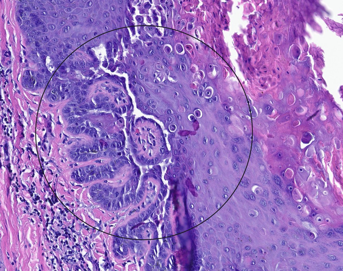
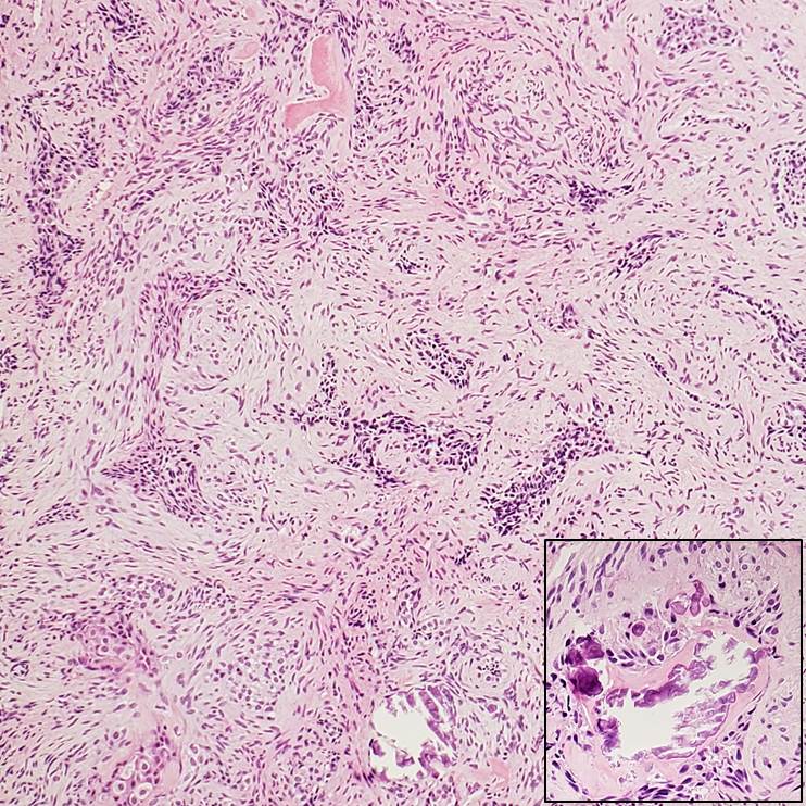
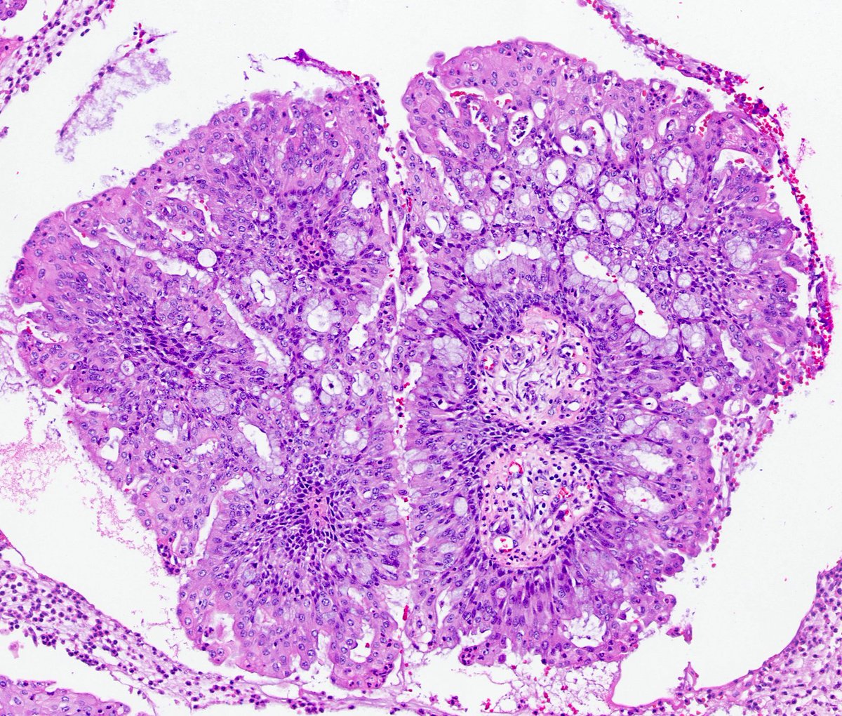
A cute mixed squamous & glandular papilloma of the lung.
These rare benign tumors show papillary cores lined by glandular epithelium admixed with squamous cells. A good reminder that not every endobronchial papillary lesion is malignant.
#PathTwitter#LungPath#MedTwitter
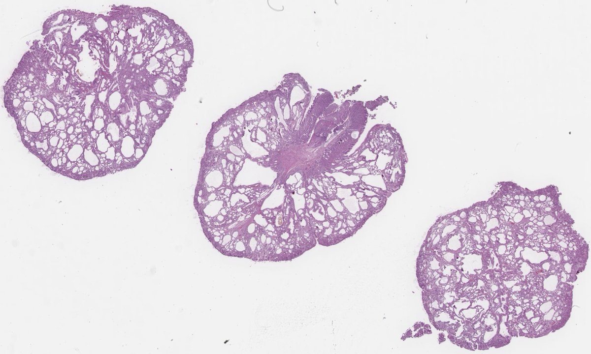
Fundic gland polyp and the screenshots are from WHO Classification of Digestive System Tumours 6th Edition online. According to it, cyctically dilated glands containing greenish material represent a characteristic feature in this type of polyp. Can anyone explain the reason?