The big one is finally out!! In this paper, we set out to provide insight into the fundamental question;
How do the individual cells from complex tissues regulate their proteomes?
Brief summary of our findings 👇
https://t.co/4dRXpGJTGn
Its fun spending probably upwards of 100 hours responding point by point to reviewers for either them not to even return the email for the revisions or respond in a few sentences that dont acknowledge any of the responses you made, its a really solid use of time!
Parallel Squared Technology Institute aims to scale the throughput of sensitive proteomics without compromising quantitative accuracy, depth or specificity.
Today, we share a step in this direction:
🔹 A workflow supporting the analysis of 1,130 single-cell samples per day (SPD) while preserving depth and quantitative accuracy.
1/
https://t.co/ygU4OhMME6
Got the results back from the first plasmid I design this morning! Anyone who has done this before would have found it trivial but to me it all felt like magic
Getting into a new field is a great way to reinvigorate your excitement and passion for science
This gets particularly interesting in single cells from the same cell type.
Working at the cell type level misses much of the action.
2/
https://t.co/49lnT3M7MX
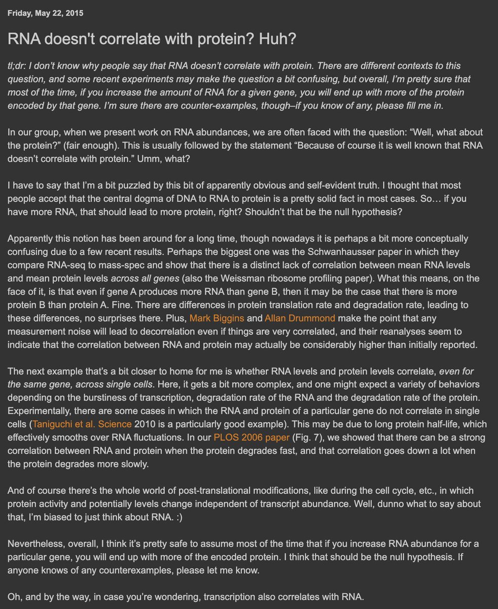
No reason to take a hard stance on this! Certain mechanisms cause regulation by translation/protein clearance in certain contexts. We aim to discover these mechanisms partly through measuring protein dynamics in single cells :)
https://t.co/PVeQSHxUaw
The piece revisits the early days, our lab's initial work alongside other groups pioneering complementary approaches, together showing that mass-spec-based single-cell proteomics was feasible & promising. The proof-of-concept set the field in motion.
2/
https://t.co/05cSIJUWuH
The piece revisits the early days, our lab's initial work alongside other groups pioneering complementary approaches, together showing that mass-spec-based single-cell proteomics was feasible & promising. The proof-of-concept set the field in motion.
2/
https://t.co/05cSIJUWuH
Cells mitigate damage to DNA in surprising ways. Using a series of CRISPRi chemogenomics screens, we identified a PRDX1-dependent iron–damage axis in the DNA damage response.
Congratulations to Tom, Abe, Shaheen, Josep and all co-authors!
https://t.co/TrtzP2T9Bw
The progress on pushing a new paradigm for multiplexing MS has been amazing. Shorter LC gradients are great but will run into fundamental limits. The creative multifaceted solutions the developed will open new doors for accessability of SCP / type of experiments that can be done
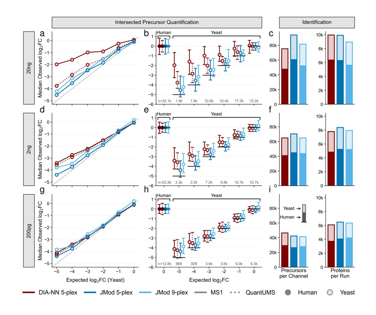
Accurately quantifying over 700K precursors in a single 9-plexDIA set of 20ng proteomes is a new milestone.
JMod achieves high quantitative accuracy over a 32-fold dynamic range of proteome spiked-in ratios across single-cell and bulk sample sizes.
1/
https://t.co/quGLcMSw9y
This analysis extended previous trends observed across cell types, where growth rate dictates the extent of control by protein degradation, to our identified gradient of growth within a cell type.
We updated our single cell in vivo metabolic labeling preprint partly thanks to constructive feedback from reviewers.
In the added analysis, we identified a gradient of proliferation within Basal epithelial cells corresponding to Krt13+ hillock cells.
https://t.co/4eA7TuPUGA
@arjunrajlab Do not waiver in your conviction! haha, I think realistically its case by case, like you have to think hard about what it would means for that specific insight to be represented in the data and how plausible it is.
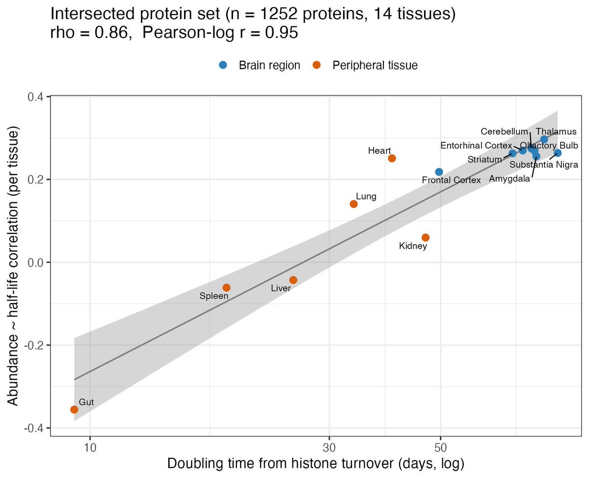
I made a mistake in the figure below and the result is actually a lot stronger (and makes much more sense, brain should clearly have slower turnover than the other tissues). Thanks @Yanshen73854711 for generating awesome data!
In this revision, we expanded our analysis across more tissue types & more model systems.
The conclusions held.
Proteomes are shaped by tissue type-specific protein degradation rates
▶️ This effect strongly depends on the cell growth rates.
🔗https://t.co/3rAvPkZuUN
@anshulkundaje@slavovLab Essentially the change was I just string grabbed for all histone proteins, but the minor variant histone are less representative of proliferation. The above plot is only using the core histones to estimate tissue turnover.