The anterior right pulmonary vein antrum, an under appreciated topography? Atrial diverticulum seen here, perhaps culprit of temperature & impedance limiting during Radiofrequency #Ablation. Imaged during unrelated, Focal Atrial #Tachycardia#Ultrasound#EPeeps credit @Ashit_EPS
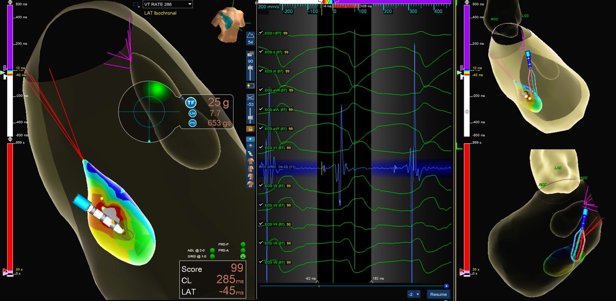
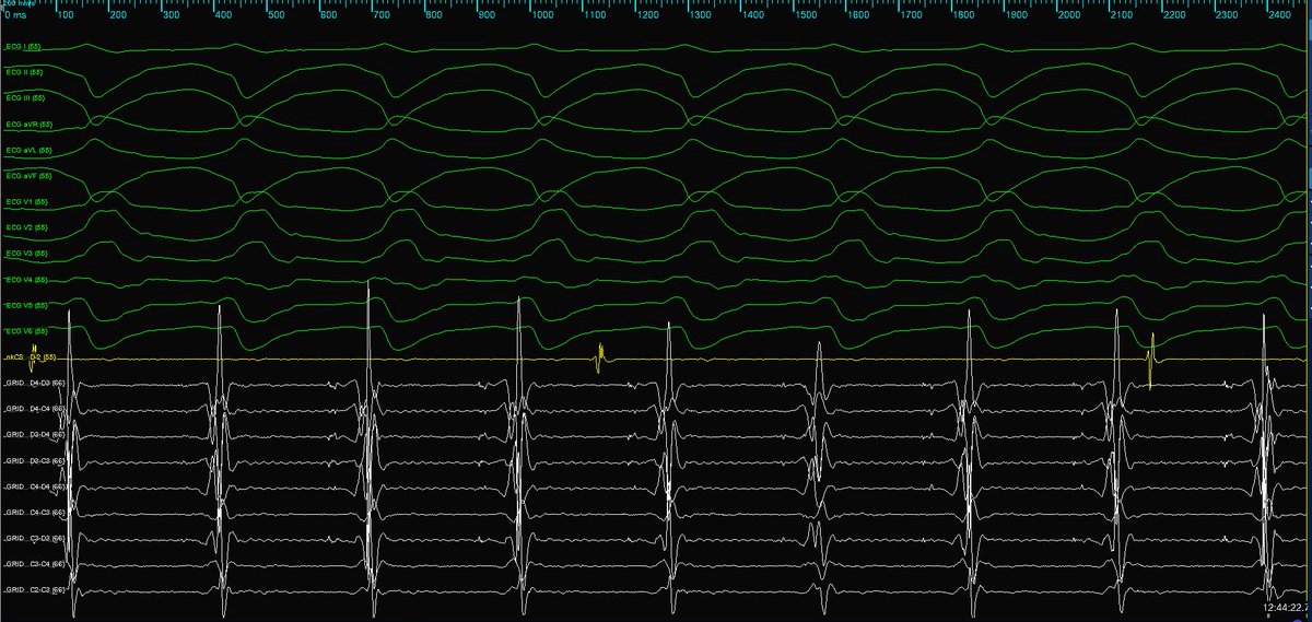
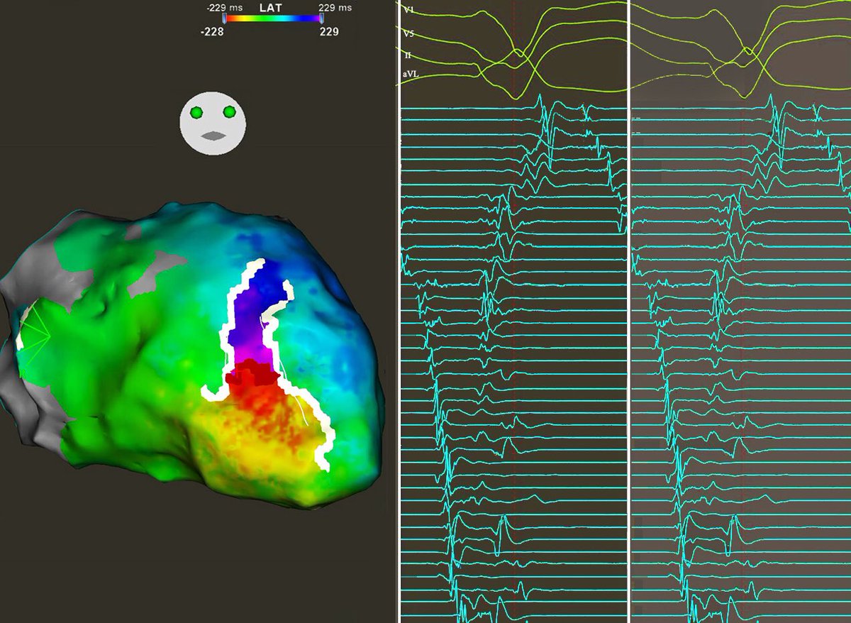
Great VT week with @BiosenseWebster! #ablateVT Inferoapical isthmus from large Cx infarction. White line is LOB in ILAM, serving as a surface barrier that forces intramural conduction. Need greater resolve of SNO zone, as it is most critical region. Great collaboration ahead!
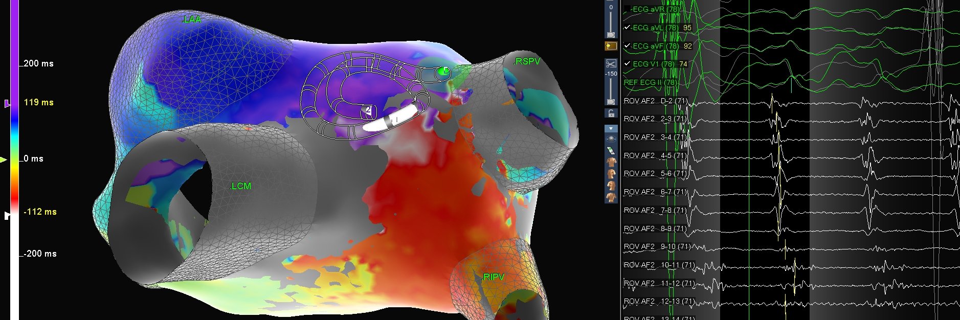
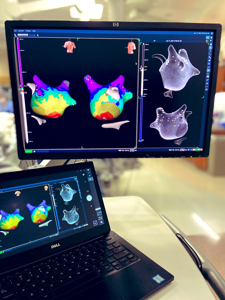
An “Atypical” Atrial Flutter disguised as #Afib revealed with strict EnSite AutoMap Stability, Correlation & Velocity discriminators. Single RF application restored sinus rhythm. #AlwaysBeMapping#AbbottAF @AbbottEP
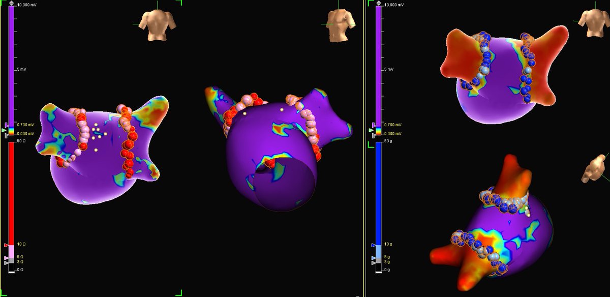
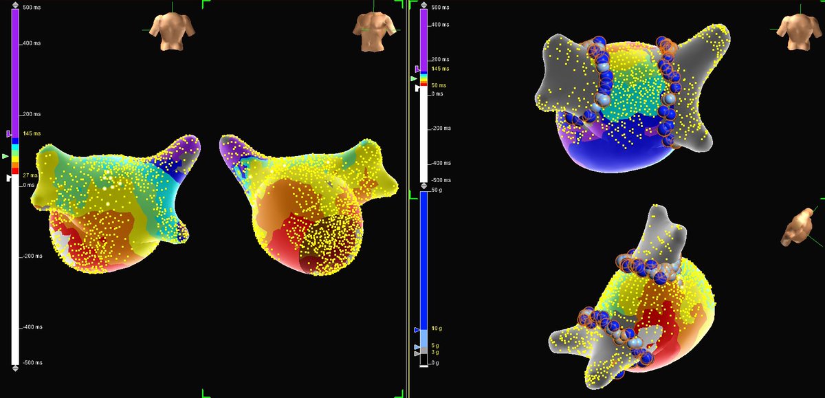
Efficient single pass isolation for this paroxysmal #AFib s/p mitral valve replacement. A notably clean surgical LAA resection, unable to visualize on ICE or geometric reconstruction. #AbbottAF @AbbottEP @TheArtOf_EP
Intrapericardial saline/air to displace the phrenic nerve - monitoring contact force sensing vector and ablation catheter tip position with ICE is helpful @drluissaenz
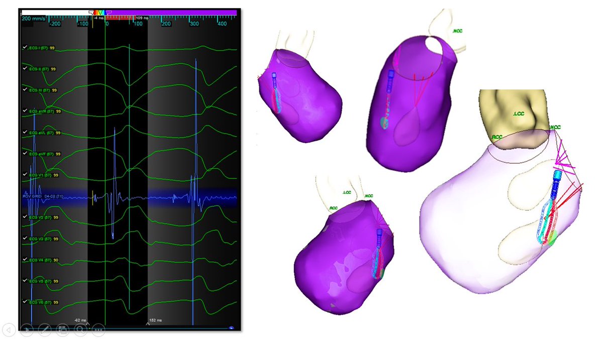
Ischemic cardiomyopathy, multiple shocks for monomorphic VT degenerating to VF. Induced with ease during SR EAM. PM papillary segmentation with PDX technique #HDGrid identified intracavitary pre-potentials #EPeeps@Ashit_EPS@B_Naz_MD https://t.co/RRLshGPB6O @AbbottGlobal
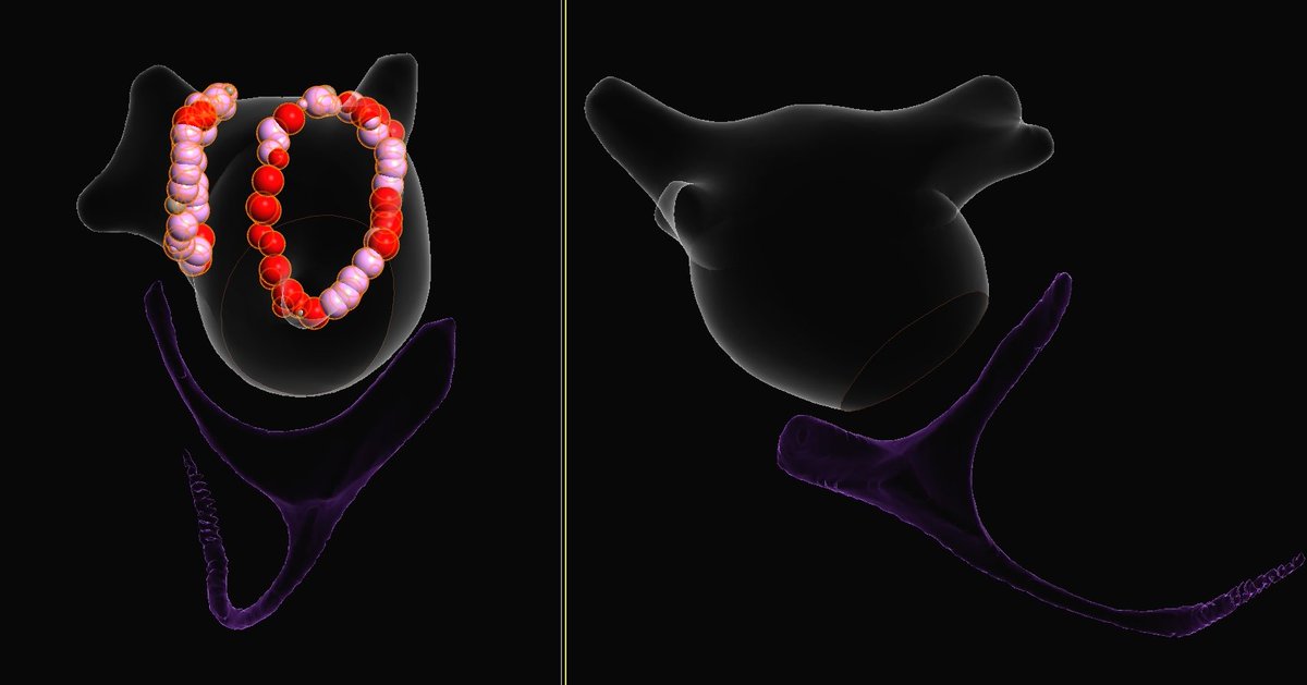
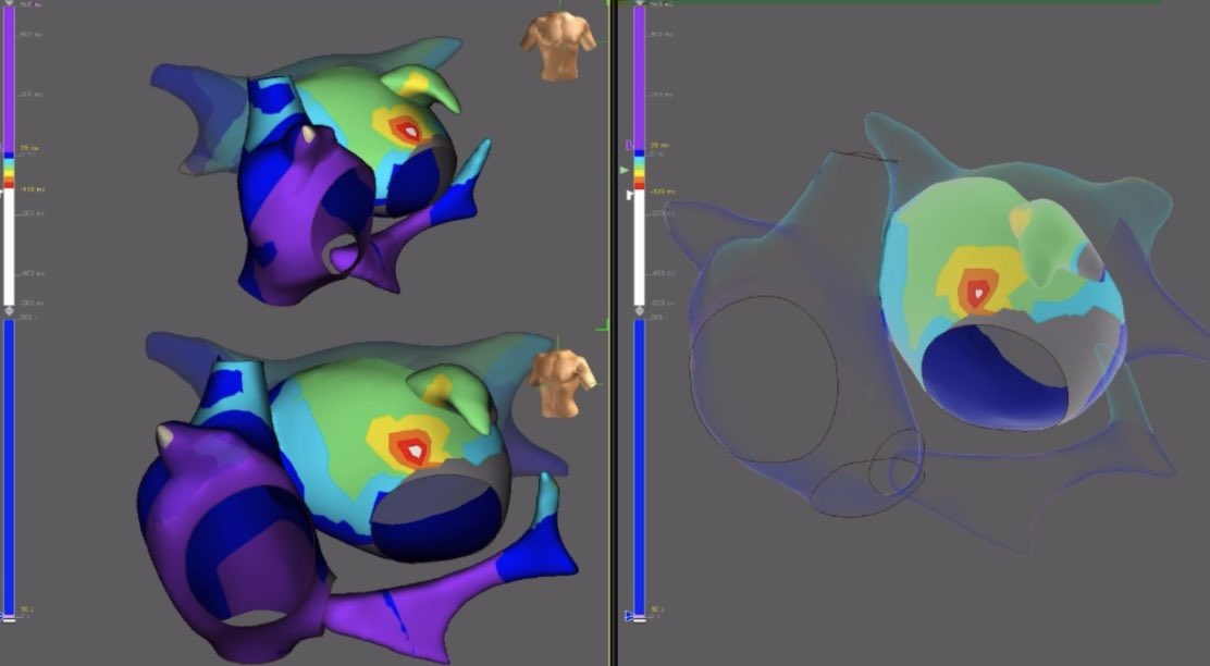
First isolated the RPVs and post wall to clean up the substrate (good tip from @Davilandre) which made interpreting the induced flutter straightforward! Terminated with first abl then anchored to prior lines. Noninducible after! @forkknifecab_EP#HDGrid#EPeeps