As of last month, I’ve officially completed my PhD! 🧬
These past few years have been incredibly rewarding and challenging in equal measure. Deep gratitude to the @ericmiskalab and to everyone who's come along the way
Just in case anyone is still here, check our latest preprint, right on time before the holiday season. We explored how transposable elements (TEs) diversify eukaryotic proteomes and found a cool case in nematode F-box genes.
https://t.co/W3HYX2khNf
Short 🧵 with highlights.
Maybe my last post here? ---> Follow the lab on 🦋🦋🦋 ! Happily retweeting new preprint: @Paul_Chammas postdoc paper @MRC_LMS , teasing apart Dux vs MERVL in the 2C-like state. Not all 2C-like cells are created equal....
The latest issue of @MolecularCell is out! Huge congrats to @josh_danac and the Tchasovnikarova lab on their #firstpublication 👏🎉➡️https://t.co/qlBq7OBvke. Read how HUSH2, a new #epigenetic regulator that works in concert with the related HUSH complex, orchestrates the immune response to invasive retroelements. @rachydoodahh @AkhilaGungi
Saying goodbye to Imke who has finished her excellent MSc abroad from the NL with us and welcoming Audrey who joins us for a postdoc! Celebrated with a 1st and 3rd place at the pub quiz 🥳🥳🥳
rnaCrosslinkOO is out in @OUPBioinfo!
https://t.co/wHxXfXYyiO
An easy to use @_R_Foundation package designed to analyse RNA cross-linking data. Raw reads to comparison of predicted structures. Data is available so please check out your favourite RNA. 1/3
Latest lab preprint! Work led by newly minted PhD Fu Xiang Quah (not on X). Using a pangenome approach to characterise structural variants (SVs) in cichlids, Fu found a substantial amount of SVs attributable to transposable elements (TEs). 🧬🐠🐠🧬
https://t.co/fdjnlI11Io
🚨New preprint alert!!🚨 I am delighted to share my first postdoc research paper on the interaction between transposable elements (TEs) and piRNAs in African cichlid fishes 🐠🐠
Link here: https://t.co/Rj4qA4YN8r
Find the highlights in 🧵
A position has become available to join @richard_durbin and his team as a Research Assistant supporting research on Malawi cichlid and Yponomeuta moth projects.
���Deadline 13 March
👉https://t.co/LwznJ5B8Dc
Our paper on the ancestral genome-assisted TE annotation method/resource is out in @CellGenomics! Enhanced annotation is now available for multiple species, including human and mouse, with ~70% increase in ancient TEs.
https://t.co/kmyUE3WGqu
Excited to say our review of genome editing in cichlid fishes, focusing on CRISPR/Cas9 tech, is now published in @RSocPublishing#OpenBiology!
Link here: https://t.co/BTsM8XTzYv
Some highlights in🧵
This paper is dense and deserves a break down.
Here you go:
There are increasing reports suggesting that a COVID-19 infection might either start or speed up neurodegenerative diseases like Alzheimer's disease (AD) and Creutzfeldt-Jakob disease (CJD).
These diseases (and others) are caused by human proteins misfolding into a structure called an "amyloid fibril." Once these fibrils form, they can cause more proteins to misfold in a chain reaction.
SARS-CoV-2 Spike Protein:
•The researchers previously found a potential mechanism where the spike protein of the SARS-CoV-2 virus (which causes COVID-19) can turn into these problematic amyloid fibrils.
•This happens when the spike protein is cleaved (or cut) by an enzyme called neutrophil elastase, which is abundant when the body is fighting a COVID-19 infection.
Main Findings:
•In their experiments, they found that these spike-protein-formed amyloid fibrils can speed up the formation of amyloid fibrils from the human prion protein (HuPrP), which is associated with CJD.
•They also tested other disease-associated amyloid fibrils to see if they had the same effect, but it seems the effect is specific to the spike-protein-formed fibrils.
•Additionally, they found that the formation of amyloid fibrils associated with Alzheimer's disease (Aβ1-42) was also sped up by these spike-protein-formed fibrils.
•They identified specific segments of the spike protein that were most efficient in causing this acceleration in different proteins.
Conclusions & Implications?
While these findings are based on in vitro experiments (meaning outside a living organism, like in a petri dish), they suggest that the spike protein's amyloid fibrils might play a role in the reported increase of CJD, AD, and potentially other neurodegenerative diseases after COVID-19.
I’ve seen some immediately claim that this means it causes Mad Cow disease.
Its important to keep in mind that while Mad Cow Disease and the potential effects of the SARS-CoV-2 spike protein both involve misfolded proteins, they are related to different types of misfolded structures (prions for Mad Cow and amyloid fibrils for the potential COVID-19 connection). However, the authors suggest a possible link between the SARS-CoV-2 spike protein and the acceleration of conditions like CJD, which is in the same family of diseases as the variant caused by Mad Cow.
Pan-mammalian epigenetic clocks are highly accurate across species, from mice to whales! 🐭🐋These clocks predict mortality risk, hinting at their value in preclinical studies. Epigenetic aging processes are evolutionarily conserved across mammals, intertwining aging and development. #Evolution #EpigeneticClocks #AgingResearch
https://t.co/nhPPVHU4Ru
Buckle up! We're in for a wild ride today. A new @NatMetabolism paper by scientists from China adds a surprising twist to the long-known FTO GWAS story.
The FTO locus (16q12.2) is the first ever GWAS locus to be associated with obesity and even after 16 yrs now, scientists appear to be scratching their heads trying to make sense of this locus. Non-coding intronic variants within FTO strongly associate with BMI, where individuals homozygous for the top risk variant weigh ~3kg more than non-carriers (https://t.co/vh4ceXVQaQ).
Since its discovery in 2007, there have been tremendous efforts to identify the causal gene(s) at this locus. Given that the risk variants are sitting right within a gene, FTO was of course the primary suspect. How do you find out if FTO has an effect on BMI? Delete it in mice and see if the animal gains weight. And that's what scientists did and found out that Fto knockout mice were stunted and lean, and the leanness was mainly due to burning too much fat (https://t.co/DkKIGLvD0P). That's great. So FTO must be the causal gene.
But then contradicting findings appeared. If you knock out the Fto only in adipose tissue (https://t.co/W5A8JwT2Hx) or globally after the animal has grown (https://t.co/uJ69bO8A9x), the mice actually gain weight!
Amidst this confusion came an even bigger one: two landmark papers, one in Nature (https://t.co/Kq3uqKrAg1) and the other in NEJM (https://t.co/Ztqf4FTOYT), said, forget about FTO, the causal genes are located far away. The FTO locus is an enhancer that folds in the 3d space and touches the promoter of distant genes IRX3 and IRX5. And deleting Irx3 in mice resulted in weight loss.
Then scientists were like, you know, a proper experiment would be not to knock out Fto or Irx3, but to delete the homologous noncoding region in mice. So, they deleted an 82 basepair-homologous region in mice and showed that without this region, the mice don't gain weight when fed with a high-fat diet, and deleting this locus increases Irx3 and Irx4 expression (https://t.co/NFFL2OQNL8). So, the causal genes are IRX3 and IRX4 then.
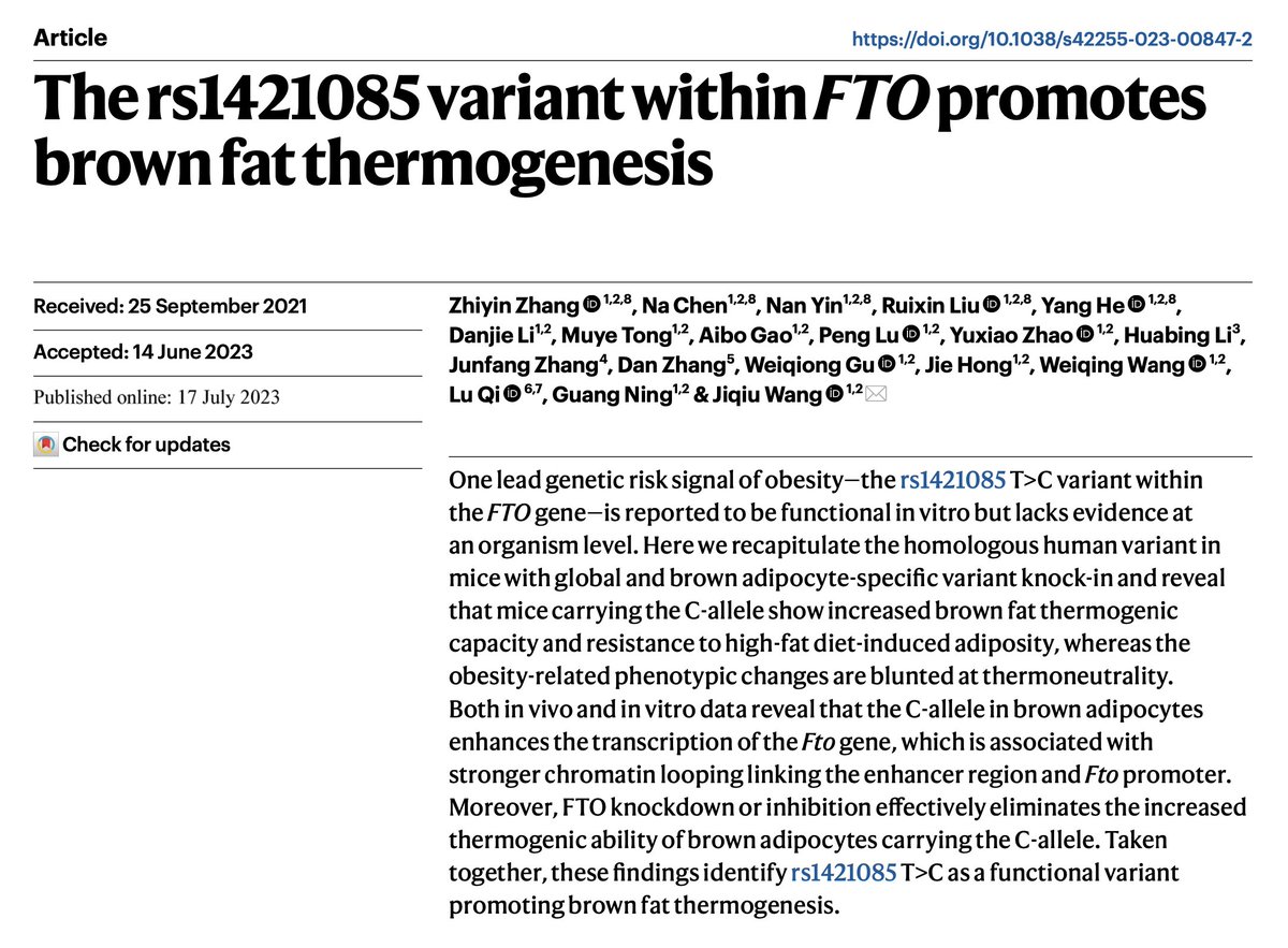
And now, in the current paper, the scientists argue, you know, the most appropriate way to study the FTO locus is to recreate the exact genetic variant in mice and study the consequences.
What did they find? The exact opposite of what was found in humans. The risk allele that increased weight in humans, decreased weight in mice. Can it be because of some off-target effects of the CRISPR experiment? No. Even if you do the knock-in in an old-fashioned way, the results are the same. The weight loss is mainly via over energy expenditure via brown adipose tissue. Wait, there is another twist.
There is an interesting difference between humans and mice. Humans have brown adipose tissue only during infancy and then lose it as they grow into adults, which isn't the case in mice. The brown adipose tissue helps mainly during cold temperatures. The experiment mice are usually housed at around 20-22 degrees Celsius, which is an ambient temperature for humans but not for mice. It's substantially cooler than mice's "thermoneutral zone (29-31 degrees)".
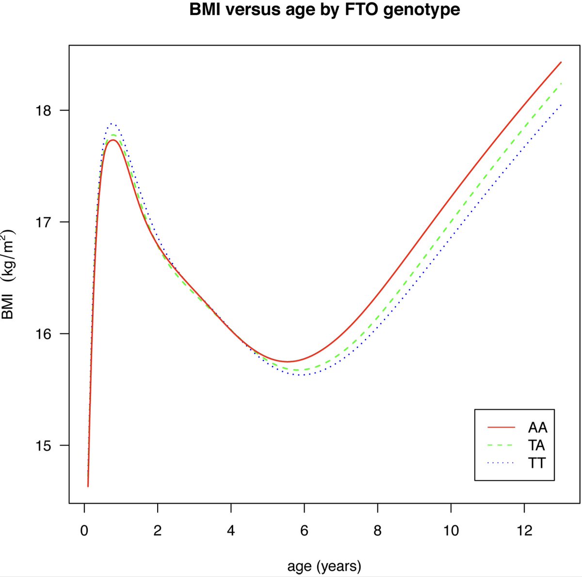
So what happens when you repeat the experiments in what might be the mice's ambient temperature--30 degrees? All the weight loss effects that were previously seen at 20-22 degrees are now blunted. So, the FTO locus effects are strongly dependent on two things: temperature and the presence of brown adipose tissue. And it turned out, in fact, it was previously shown in humans that the FTO variant has an age-dependent effect. It lowers the body weight in infants and then increases the body weight in adults, which aligns with the current finding. (https://t.co/S7iKVj0p5d)
Overall, the current paper is an impressive work and will stand as a landmark in the long twisty road of FTO story. But above all, this paper is a remarkable example to show case the challenges behind translating mice physiology to human physiology.
https://t.co/i8IIr2bSFc
Excited to share our new paper hot off the press @NatureComms on #Genomics of cold adaptations in the #Antarctic#notothenioid fish radiation. A culmination of years of work with a great team of collaborators
@SangerToL @Naturalis_Sci
@GeneticsCam
https://t.co/5Bl6qAETH0
Our latest preprint! David Jordan surveys the phenotypic space of growth & development of C. elegans in different genetic & environ. contexts, to explore the principles governing the emergence of robustness and flexibility in complex biological systems. https://t.co/oa7iSVShL8
It is finally out (in a peer-reviewed journal)! Our contribution to teasing out human placental gene regulation by #ERVs. Led by the awesome @neverlethetruth.
https://t.co/Ha2JIFHDy0